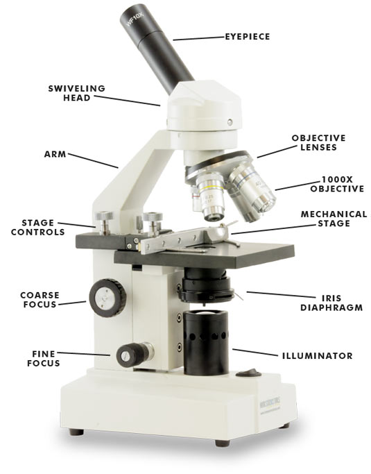
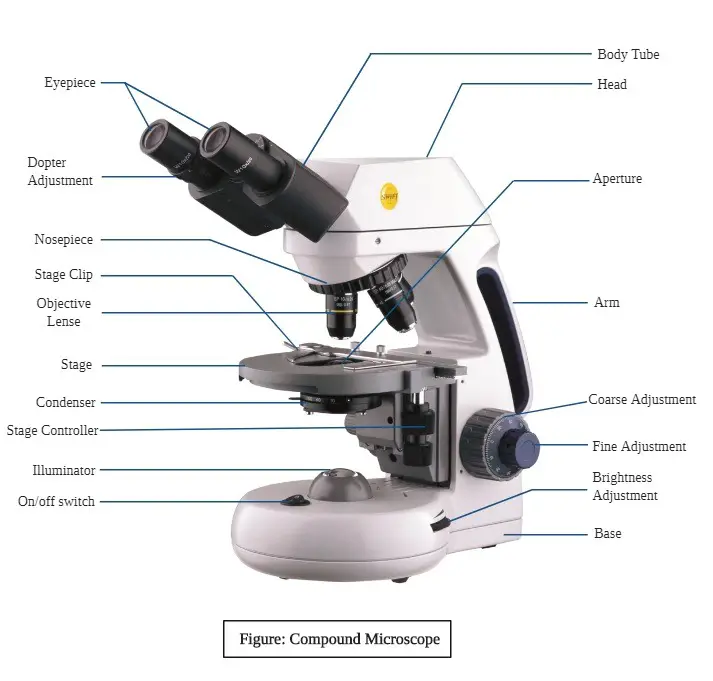
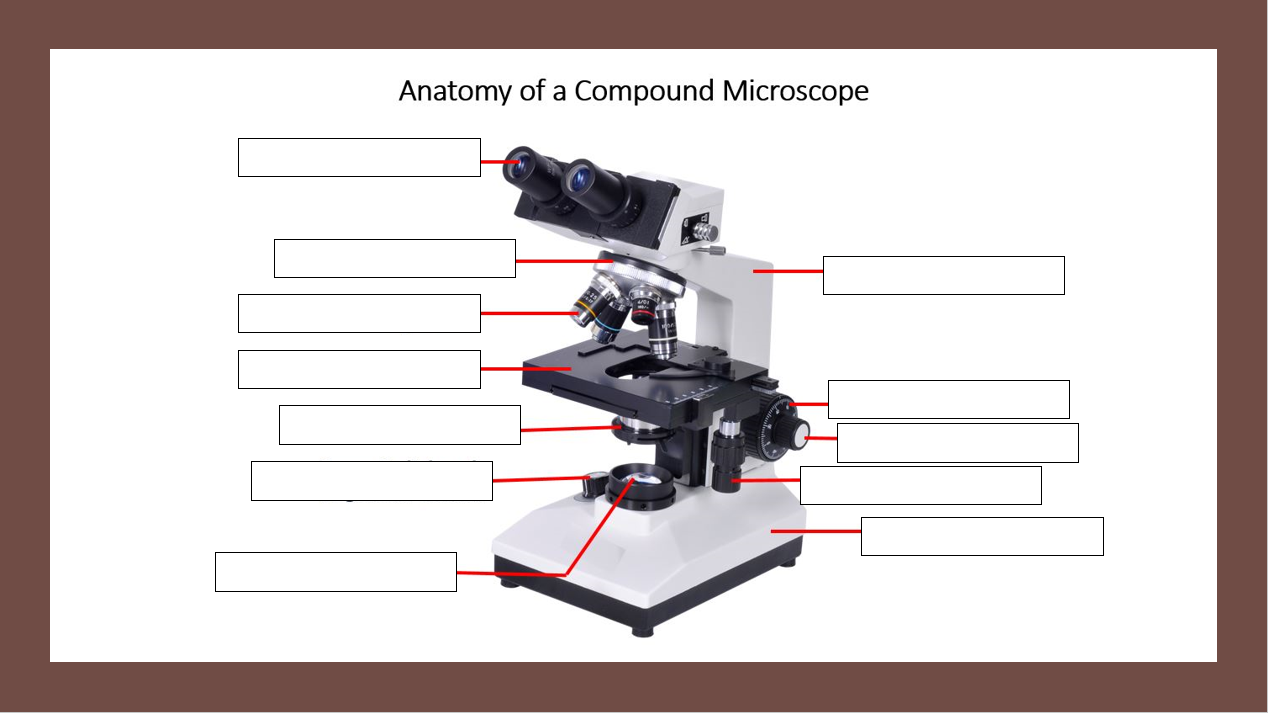
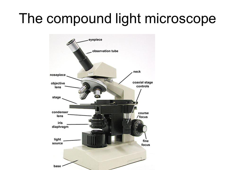
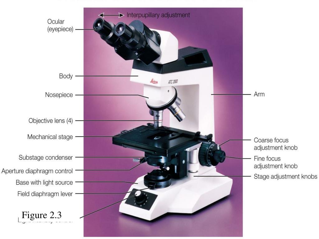
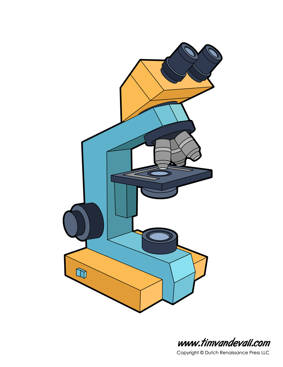
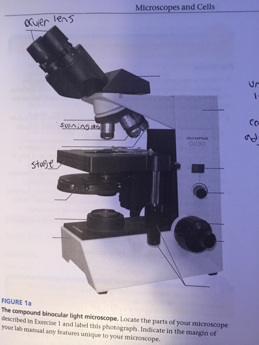
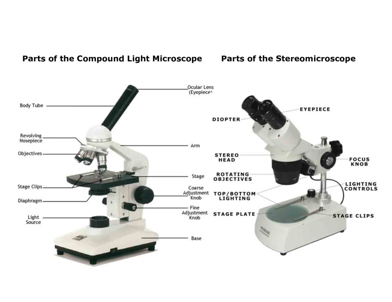
39 compound light microscope diagram
In order to view small objects with a compound microscope, the object, or ... Preparing a wet mount of a specimen is the technique typically used to view plant and animal cells using a microscope. This page provides step by step instructions on slide preparation as well as videos at the bottom of page. How to Prepare a Wet Mount Slide. Page last updated: 1/2016. Step Five - If the specimen is ... Anatomy Of A Microscope Microscope Illumination Olympus Life. Microscope Parts And Functions. Compound Microscope Diagram Worksheet Printable Worksheets And. Compound Light Microscope Diagram. 1581327960000000. Microscope Drawing And Label At Paintingvalley Com Explore. Solved Part D Associate The Functions Of The Compound Lig.
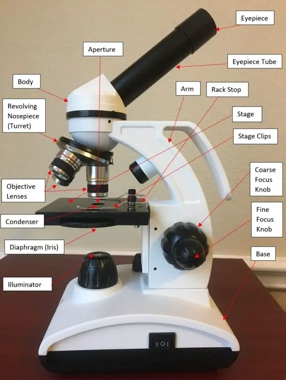
Aperture. Illuminator. Condenser. Diaphragm. Parts of a Compound Microscope. Video: Parts of a compound Microscope with Diagram Explained. As a side note, the microscope used in this post is a great entry level or beginner microscope if you are trying to get someone interested in microscopes, microbiology, or science in general.
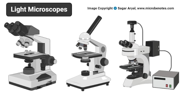
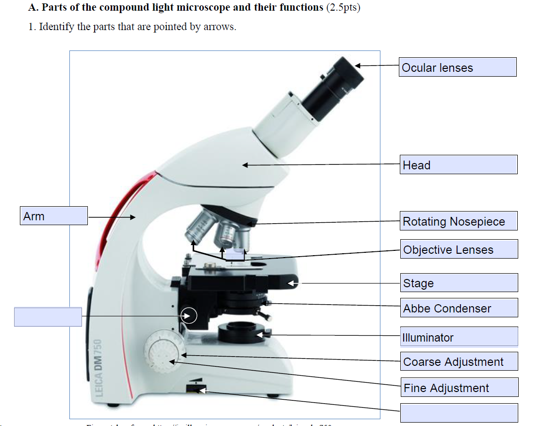
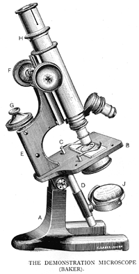
Compound light microscope diagram
Using a light microscope, it is just barely possible to see tiny green granules—which were named grana. With ... 2 in a four-carbon compound, which is why the process is called C 4 photosynthesis. The four-carbon compound is then transported to the bundle sheath chloroplasts, where it drops off CO 2 and returns to the mesophyll. Bundle sheath chloroplasts do not carry out the light reactions ... The light microscope can extend our ability to see detail by 1000 times, so that we can see objects as small as 0.1 micrometer (um) or 100 nanometers (nm) in diameter. The transmission electron microscope extends this capability to objects as small as 0.5 nm in diameter, 1/200,000th the size of objects that are visible to the naked eye. Without Microscope Parts and Functions With Labeled Diagram and Functions How does a Compound Microscope Work?. Before exploring microscope parts and functions, you should probably understand that the compound light microscope is more complicated than just a microscope with more than one lens.. First, the purpose of a microscope is to magnify a small object or to magnify the fine details of a larger ...
Compound light microscope diagram. Most compound light microscopes will contain three to four objective lenses that can be rotated over the slide. Sometimes these lenses are just called ... 26.10.2021 · A light microscope uses focused light and lenses to magnify a specimen, usually a cell. In this way, a light microscope is much like a telescope, except that instead of … Compound Microscope – Diagram (Parts labelled), Principle and Uses. ... Also called as binocular microscope or compound light microscope, it is a remarkable magnification tool that employs a combination of lenses to magnify the image of a sample that is not visible to the naked eye. within the eukaryotic cell. The microscope you will be using is a compound microscope which utilizes two lenses or lens systems to enlarge the object being viewed. The ocular or eyepiece is the lens near your eye while the objective is the lens located on the revolving nosepiece. Your microscope has two oculars, or binocular vision. The object ...
Essential Parts of Compound Microscope: · (i) Lenses: · (ii) Adjustment of Objective Lens: · (iii) Stage: · (iv) Mirror: · (v) Sub-Stage Diaphragm: · (vi) Sub-Stage ... A compound light microscope is a type of light microscope that uses a compound lens system, meaning, it operates through two sets of lenses to magnify the image of a specimen. It’s an upright microscope that produces a two-dimensional image and has a higher magnification than a stereoscopic microscope. 18 Nov 2020 — Parts of a Compound Microscope · Eyepiece (ocular lens) with or without Pointer: The part that is looked through at the top of the compound ... Prostate cancer is cancer of the prostate.The prostate is a gland in the male reproductive system that surrounds the urethra just below the bladder. Most prostate cancers are slow growing. Cancerous cells may spread to other areas of the body, particularly the bones and lymph nodes. It may initially cause no symptoms. In later stages, symptoms include pain or difficulty urinating, blood in the ...
The compound light microscope is an instrument containing . two lenses, which magnifies, and a variety of . knobs to resolve (focus) the picture. Because it uses more than one lens, it is sometimes called the compound microscope in addition to being referred to as being a light microscope. In this lab, we will learn about the proper use and handling of the microscope. Instructional Objectives ... 18 Jul 2021 — Devised with a system of combination of lenses, a compound microscope consists of two optical parts, namely the objective lens and the ... Start studying Compound Microscope Diagram. Learn vocabulary, terms, and more with flashcards, games, and other study tools. F. LIGHT SOURCE Projects light UPWARDS through the diaphragm, the SPECIMEN, and the LENSES H. DIAPHRAGM Regulates the amount of LIGHT on the specimen E. STAGE Supports the SLIDE being viewed K. ARM Used to SUPPORT the B. NOSEPIECE microscope when carried Holds the HIGH- and LOW- power objective LENSES; can be rotated to change MAGNIFICATION.
Optical components of a compound microscope — The head includes the upper part of the microscope, which houses the most critical optical components, ...
Q. The diagram below represents a hydra as viewed with a compound light microscope.If the hydra moves toward the right of the slide preparation, which diagram best represents what will be observed through the microscope?
Microscope Parts and Functions With Labeled Diagram and Functions How does a Compound Microscope Work?. Before exploring microscope parts and functions, you should probably understand that the compound light microscope is more complicated than just a microscope with more than one lens.. First, the purpose of a microscope is to magnify a small object or to magnify the fine details of a larger ...
The light microscope can extend our ability to see detail by 1000 times, so that we can see objects as small as 0.1 micrometer (um) or 100 nanometers (nm) in diameter. The transmission electron microscope extends this capability to objects as small as 0.5 nm in diameter, 1/200,000th the size of objects that are visible to the naked eye. Without

Compound Light Microscope Total Magnification Power Of The Eyepiece 10x Multiplied By Objective Lenses Determines Total Magnification
Using a light microscope, it is just barely possible to see tiny green granules—which were named grana. With ... 2 in a four-carbon compound, which is why the process is called C 4 photosynthesis. The four-carbon compound is then transported to the bundle sheath chloroplasts, where it drops off CO 2 and returns to the mesophyll. Bundle sheath chloroplasts do not carry out the light reactions ...

Monday 10 19 15 Aim How Do The Parts Of The Compound Light Microscope Work Do Now Observe The Picture Displayed And Label The Parts Of The Compound Ppt Video Online Download













0 Response to "39 compound light microscope diagram"
Post a Comment