40 gel electrophoresis labeled diagram
Written By Pelvic Diagram Saturday, September 5, 2020. Edit. Gel Electrophoresis Diagram. Gel electrophoresis is a technique used to separate mixtures like DNA and proteins. Gel electrophoresis uses a gel (like gelatin) and an electric field is put through the gel. The results of gel electrophoresis are shown below with four different strands of dna labeled which strands of dna is the shortest - 4529417
Electrophoresis (With Diagram) The term electrophoresis describes the migration of a charged particle under the influence of an electrical field. Many important biomolecules — such as peptide, proteins nucleotides and nucleic acids — possess ionisable groups and, therefore, at any given pH, exist in solution as electrically charged species ...

Gel electrophoresis labeled diagram
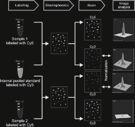
today we'll be talking about gel electrophoresis what is gel electrophoresis you might ask well it's a lab technique usually used in the biochemistry lab for separating out DNA or proteins based on their size and let's talk about how it works so first you need to have the gel this can be made out of different kinds of substances such as agarose and polyacrylamide both of which I'll discuss ... Fluorescence two-dimensional difference gel electrophoresis ... The experimental design recommended by the CyDye manufacturer uses Cy3 to label a pooled standard and the preparative gel, and Cy5 to label the pooled or test samples, because there is no Cy2-conjugated saturation dye available. Gel electrophoresis: A laboratory technique used to separate molecules, such as DNA, RNA and proteins, according to their size and charge. DNA: Deoxyribonucleic acid (DNA) is a molecule that contains the instructions needed for an organism to develop and function.
Gel electrophoresis labeled diagram. 2. The advent of capillary gel electrophoresis. Capillary electrophoresis was introduced almost 30 years ago by Jorgensen as a new and automated alternative to slab gel electrophoresis [].As early as 1989, Bob Brownlee and coworkers introduced the first commercial instrument with on column UV/VIS detection, automatic injection and computerized data analysis for rapid, high-resolution CE ... Sort and measure DNA strands by running your own gel electrophoresis experiment. Click to unmute. See how gel electrophoresis is used in forensics. Can DNA Demand a Verdict? Try it Yourself. How to Build an Electrophoresis Chamber (PDF) Colorful Electrophoresis. Funding. Funding provided by grant 51006109 from the Howard Hughes Medical ... Based on the data they collected using gel electrophoresis, label the branching tree diagram below. Write the letters A, B, and C, to represent the possible evolutionary relationships between species A, B, and C Using the circle provided, construct a labeled diagram of the restriction map of the plasmid. Explain how you developed your map. (b) Describe how: ... then separated with electrophoresis, as shown. RESULTS OF GEL ELECTROPHORESIS Kilobase 100 FxoRI + Molecular Weight Standards .
Protein Electrophoresis Guide. A guide to polyacrylamide gel electrophoresis and protein detection, including theory, product selection, protocols, and more. 2-D Electrophoresis Workflow How-To Guide. This guide describes the experimental methods and tools used in 2-D electrophoresis and proteomics research. Diagram of agarose gel setup, for agarose gel electrophoresis. (Figure by MIT OpenCourseWare.) Today you will separate DNA fragments using an agarose matrix. Agarose is a polymer that comes from seaweed and if you've ever made Jell-O™, then you already have all the skills for pouring an agarose gel. Start studying Gel Electrophoresis. Learn vocabulary, terms, and more with flashcards, games, and other study tools. the solidified gel. Remove the tape from the ends of the gel tray. 6. Place gel into electrophoresis unit. Add 150 ml 1X TBE buffer to completely fill the box and to cover the top gel surface with about 2 mm of buffer. NOTE: At this point the gel box can be covered and left until the next day if necessary 7.
Gel Electrophoresis. Lane 1: DNA Ladder. Lane 2: Undigested plasmid A. Lane 3: Completely digested plasmid A. Lane 4: Digested PCR product (or DNA Fragment). Lane 5: PCR Product (with a faint primer dimer band). Lane 6: Genomic DNA. The white arrows indicate the bands that you want to excise. Question: 4. Use the shapes/ text box features in Word to draw and label a diagram of a gel electrophoresis unit. Make sure to include: a. The wells (1 pt.) b. The anode and cathode (and their charge) (1 pt.) A ladder with fragments at 10, 50, 100, and 500 base pairs (bp) (1 pt.) d. Sample 1: with fragments at 75 bp and 75 bp (1 pt.) e. Gel electrophoresis is a technique used to separate DNA fragments according to their size. DNA samples are loaded into wells (indentations) at one end of a gel, and an electric current is applied to pull them through the gel. DNA fragments are negatively charged, so they move towards the positive electrode. Because all DNA fragments have the ... The following diagram demonstrates two Serum Protein Electrophoresis (SPEP) patterns from different patients. The pattern on the left is from a normal, healthy patient. The pattern on the right is from a patient with a particular disorder. What abnormality is suggested by the pattern on the right?
2d Dige Saturation Labeling For Minute Sample Amounts Difference Gel Electrophoresis Dige Methods And Protocols Page 91
(a) Using the circle provided, construct a labeled diagram of the restriction map of the plasmid. Explain how you developed your map. Construct a labeled map and explain (3 points maximum) H H E E 40 20 30 10 H E 40 20 30 10 E H E = EcoRI Restriction Point H = HaeIII Restriction Point

Indesign Labeling Annotating Pcr Gel Pictures Advanced Tutorial Part 12 Youtube
Agarose Gel Electrophoresis. Agarose gel electrophoresis separates DNA fragments according to their size. Typically, a DNA molecule is digested with restriction enzymes, and the agarose gel electrophoresis is used as a diagnostic tool to visualize the fragments.An electric current is used to move the DNA molecules across an agarose gel, which is a polysaccharide matrix that functions as a sort ...
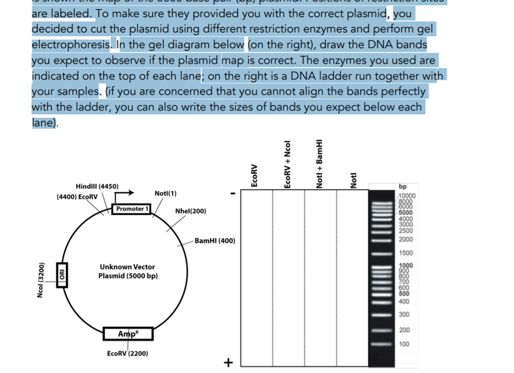
Solved You Received A Vector Plasmid From A Laboratory Next Chegg Com
Gel electrophoresis A technique used to separate DNA fragments and other macromolecules by size and charge. Key points: Gel electrophoresis is a technique used to separate DNA fragments according to their size. DNA samples are loaded into wells (indentations) at one end of a gel, and an electric current is applied to pull them through the gel. DNA fragments are negatively charged, so they move ...
1
Draw a neat labelled diagram of a typical agarose gel electrophoresis. Easy. Answer. In agarose gel electrophoresis, agarose is used as a matrix. The sample is added in the slot and current is applied to it. The smaller molecules move faster and the larger molecules are retarded. In this method, separation is based on charge and size of the ...

Agarose Gel Electrophoresis Results Analysis Video Lesson Transcript Study Com
Gel electrophoresis is a type of biotechnology that separates molecules based on their size to interpret an organism's DNA. An enzyme is used to separate a strand of DNA from a source and the DNA is suspended in a dye. Then, the dye is applied to a negatively-charged gel on one side of a sheet.
Bio 140 Lab Gel Electrophoresis Prelab Quiz Diagram Quizlet
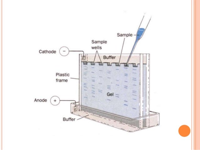
Gel electrophoresis labeled diagram. An enzyme is used to separate a strand of dna from a source and the dna is suspended in a dye. A lot of expertise and experience are required for interpreting gel electrophoresis. Electrophoresis units are available for running either vertical or horizontal gel system.

Draw A Diagram Of A Typical Agarose Gel Electrophoresis Showing Migration Of Undigested
Gel electrophoresis employs a buffer system, a medium which is a gel and a source of direct current (Fig. 6). Samples having DNA fragments are applied on the gel and current is passed through the system for an appropriate time. Different DNA fragments move up to different distances on the gel depending on their charge to mass ratio. The heavier ...

Gel Electrophoresis To Determine Genotype Youtube
Gel Electrophoresis Steps. The broad steps involved in a common DNA gel electrophoresis protocol: 1. Preparing the samples for running. The DNA is isolated and preprocessed (e.g. PCR, enzymatic digestion) and made up in solution with some basic blue dye to help visualize the movement of the sample through the gel. 2.

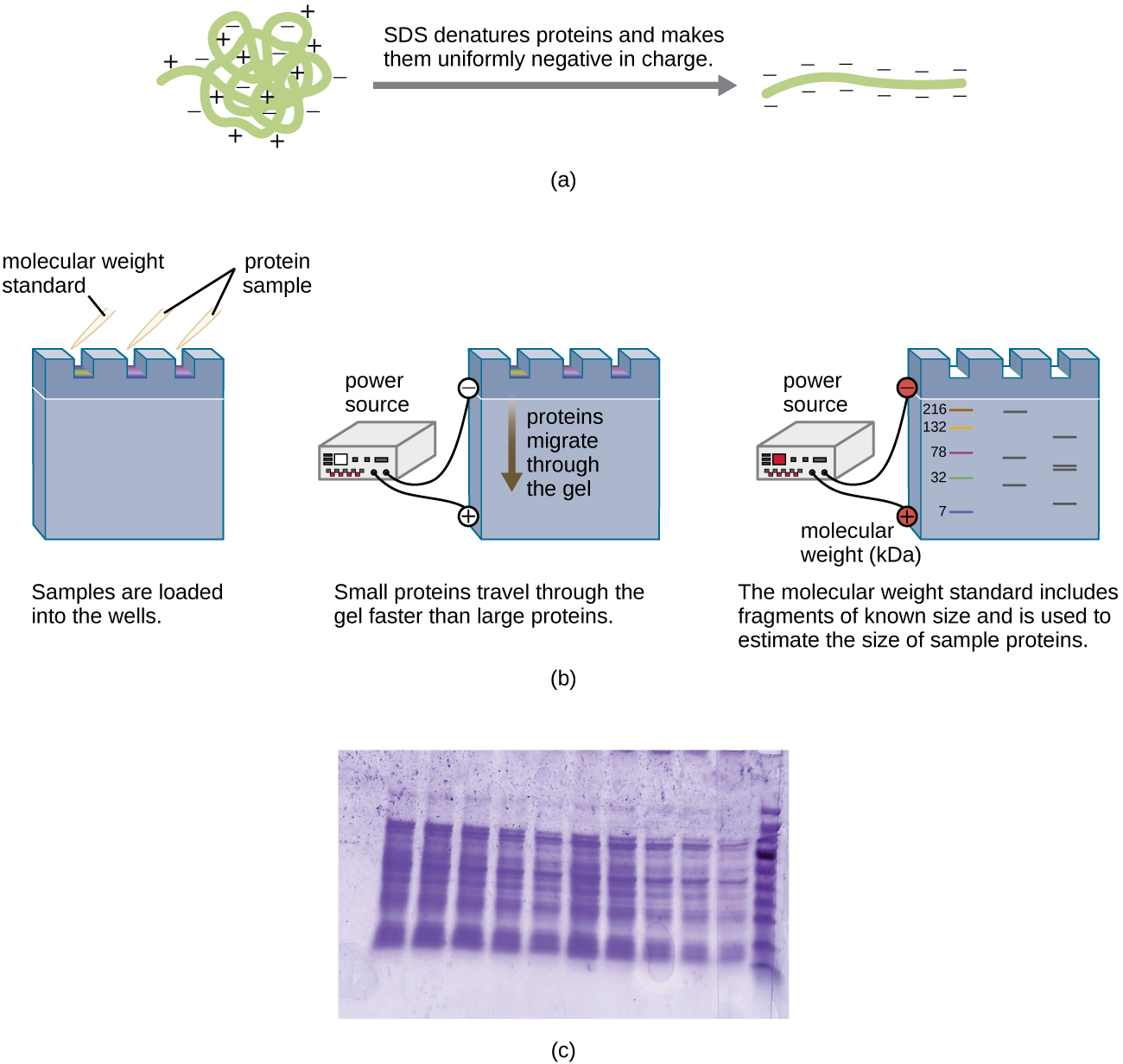
Visualizing And Characterizing Dna Rna And Protein Microbiology
Agarose gel electrophoresis is the most effective way of separating DNA fragments of varying sizes ranging from 100 bp to 25 kb 1.Agarose is isolated from the seaweed genera Gelidium and Gracilaria, and consists of repeated agarobiose (L- and D-galactose) subunits 2.During gelation, agarose polymers associate non-covalently and form a network of bundles whose pore sizes determine a gel's ...

Gel Electrophoresis Types Principles Instrumentation And Applications Microbiology Notes
On the gel diagram at the right, show how you believe these fragments will sort out during electrophoresis. • Label each fragment with its correct number of base pairs. 11 Staining DNA with Fast Blast DNA Stain (Laboratory Procedure) Consideration 3.
12 2 Visualizing And Characterizing Dna Biology Libretexts
Gel electrophoresis: A laboratory technique used to separate molecules, such as DNA, RNA and proteins, according to their size and charge. DNA: Deoxyribonucleic acid (DNA) is a molecule that contains the instructions needed for an organism to develop and function.

What Is A Dna Fingerprint Facts Yourgenome Org
Fluorescence two-dimensional difference gel electrophoresis ... The experimental design recommended by the CyDye manufacturer uses Cy3 to label a pooled standard and the preparative gel, and Cy5 to label the pooled or test samples, because there is no Cy2-conjugated saturation dye available.

What Are The Steps Of Gel Electrophoresis
today we'll be talking about gel electrophoresis what is gel electrophoresis you might ask well it's a lab technique usually used in the biochemistry lab for separating out DNA or proteins based on their size and let's talk about how it works so first you need to have the gel this can be made out of different kinds of substances such as agarose and polyacrylamide both of which I'll discuss ...
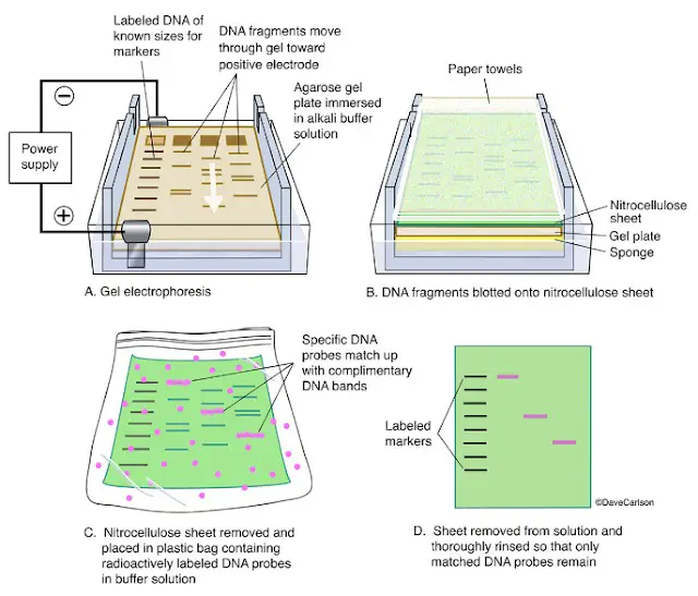
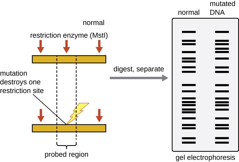
Southern Blotting Technique Principle Procedure Importance

Two Dimensional Gel Electrophoresis An Overview Sciencedirect Topics

Sds Gel Electrophoresis Of 35 S Methionine And 35 S Cystine Labeled Download Scientific Diagram

16 Dna Electrophoresis Illustrations Clip Art Istock

Evaluation Of Pcr Labeled Probes By Agarose Gel Electrophoresis On Download Scientific Diagram
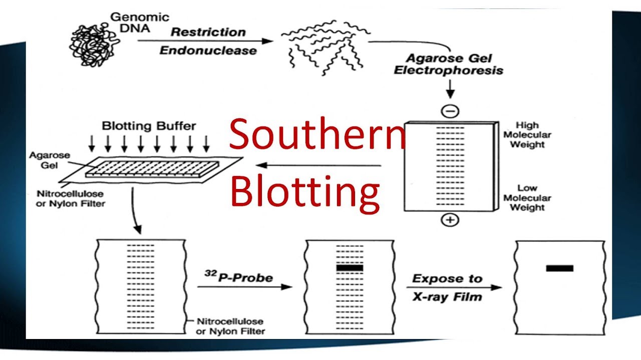
Southern Blotting Mybiosource Learning Center

Gel Electrophoresis Diagram Flashcards Quizlet

Gel Electrophoresis Bioninja

Agarose Gel Electrophoresis Principle Procedure Results Microbe Online
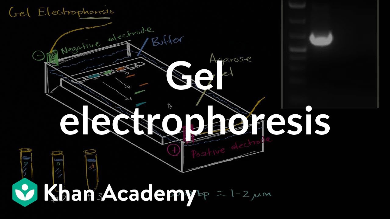
Gel Electrophoresis Video Biotechnology Khan Academy

Quantitative Protein Profiling Using Two Dimensional Gel Electrophoresis Isotope Coded Affinity Tag Labeling And Mass Spectrometry Molecular Cellular Proteomics

Addgene Protocol How To Run An Agarose Gel

Activity 2 Gel Electrophoresis Of Dyes

Electrophoresis And Blotting Of Dna Tamber Major Reference Works Wiley Online Library
1

Pin On Genetics

What Is Gel Electrophoresis Facts Yourgenome Org

Electrophoresis Gel Stock Illustrations 52 Electrophoresis Gel Stock Illustrations Vectors Clipart Dreamstime
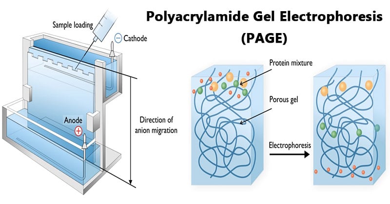
Polyacrylamide Gel Electrophoresis Page Instrumentation Microbe Notes
12 2 Visualizing And Characterizing Dna Biology Libretexts
1

Nucleic Acids And Chromatin 2 4 Analysis Of Nucleic Acids By Electrophoresis And Hybridisation Openlearn Open University S377 1
Agarose Gel Electrophoresis Of Labeled Dna In Which The Same Gel Is Download Scientific Diagram

Two Dimensional Fluorescence Difference Gel Electrophoresis For Comparative Proteomics Profiling Semantic Scholar
Gel Electroporosis
Agarose Gel Electrophoresis Age Image Of The Pcr Products A With The Download Scientific Diagram
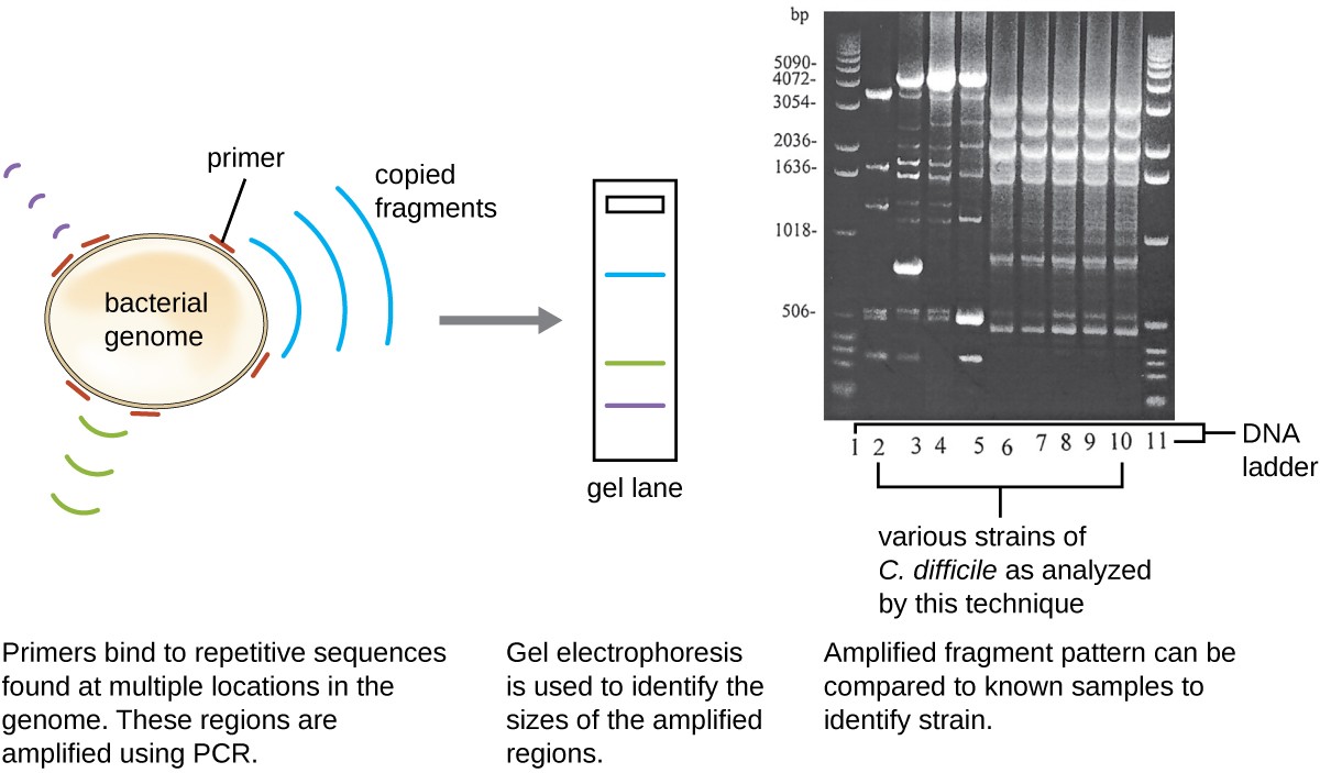
Visualizing And Characterizing Dna Rna And Protein Microbiology
0 Response to "40 gel electrophoresis labeled diagram"
Post a Comment