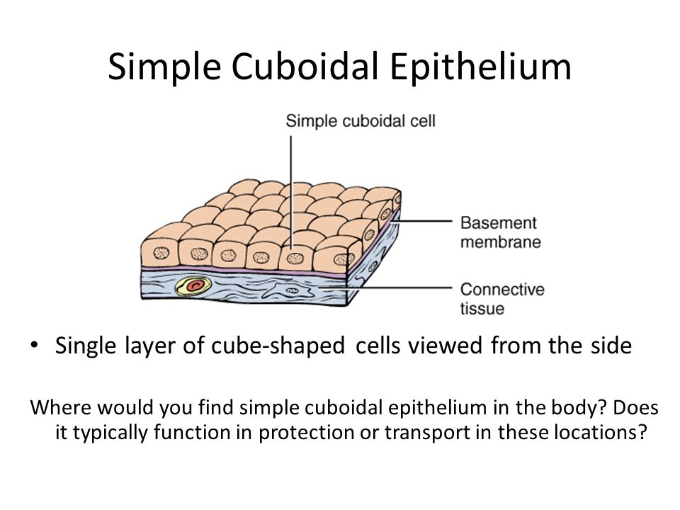
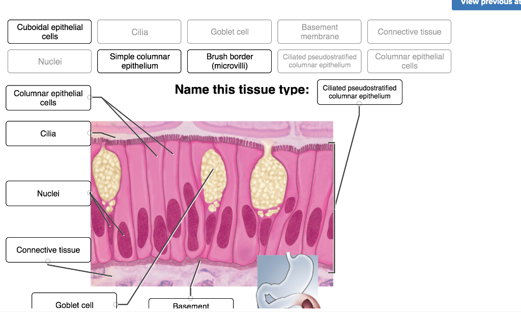
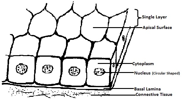
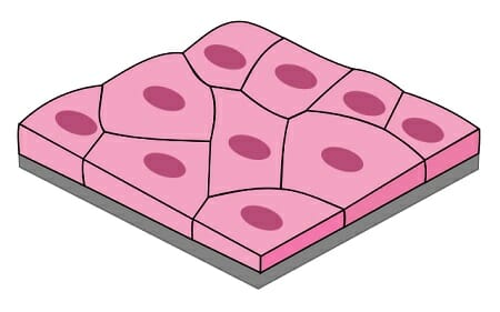
40 simple squamous epithelium labeled diagram
Which of the labeled layers in the diagram of the arterial wall is composed of a simple squamous epithelium, a basement membrane and a layer of elastic tissue? a) A b) B c) C d) A and B e) A, B, and C. a) A . Which labeled structure in the figure is a metarteriole? a) A b) B c) D d) F e) E. b) B. A blockage to one or both of the inferior phrenic veins will cause a backup of …
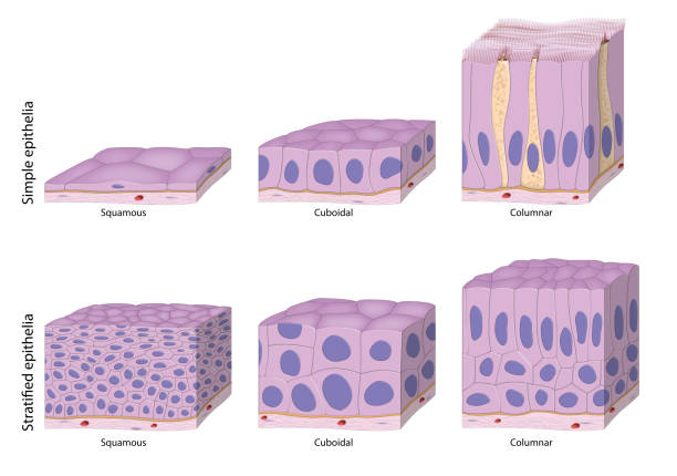
The simple in the term simple squamous epithelium means there is one layer of flat-shaped (Squamous) cells in simple squamous epithelial tissue. Simple squamous epithelial tissue is only composed...
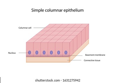
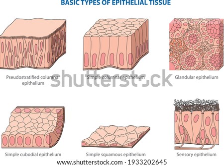
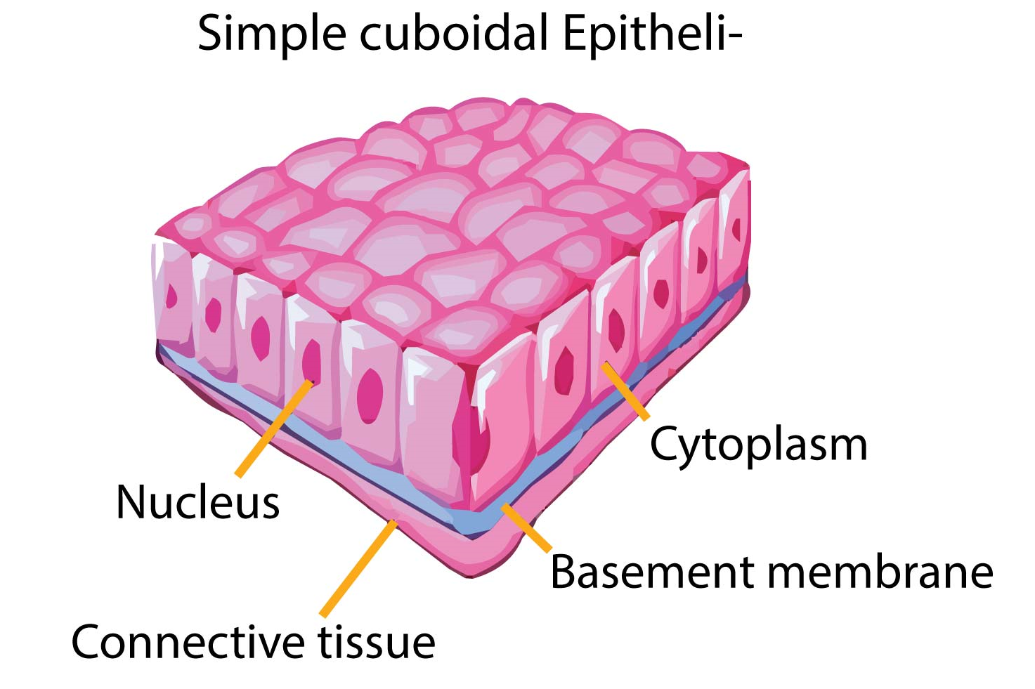
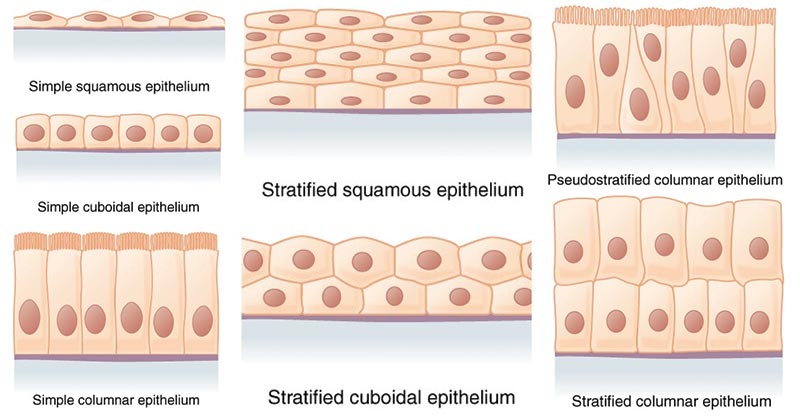
Simple Columnar Epithelium: A Labeled Diagram and Functions Epithelium is a tissue that lines the internal surface of the body, as well as the internal organs. Simple epithelium is one of the types of epithelium that is divided into simple columnar epithelium, simple squamous epithelium, and simple cuboidal epithelium.
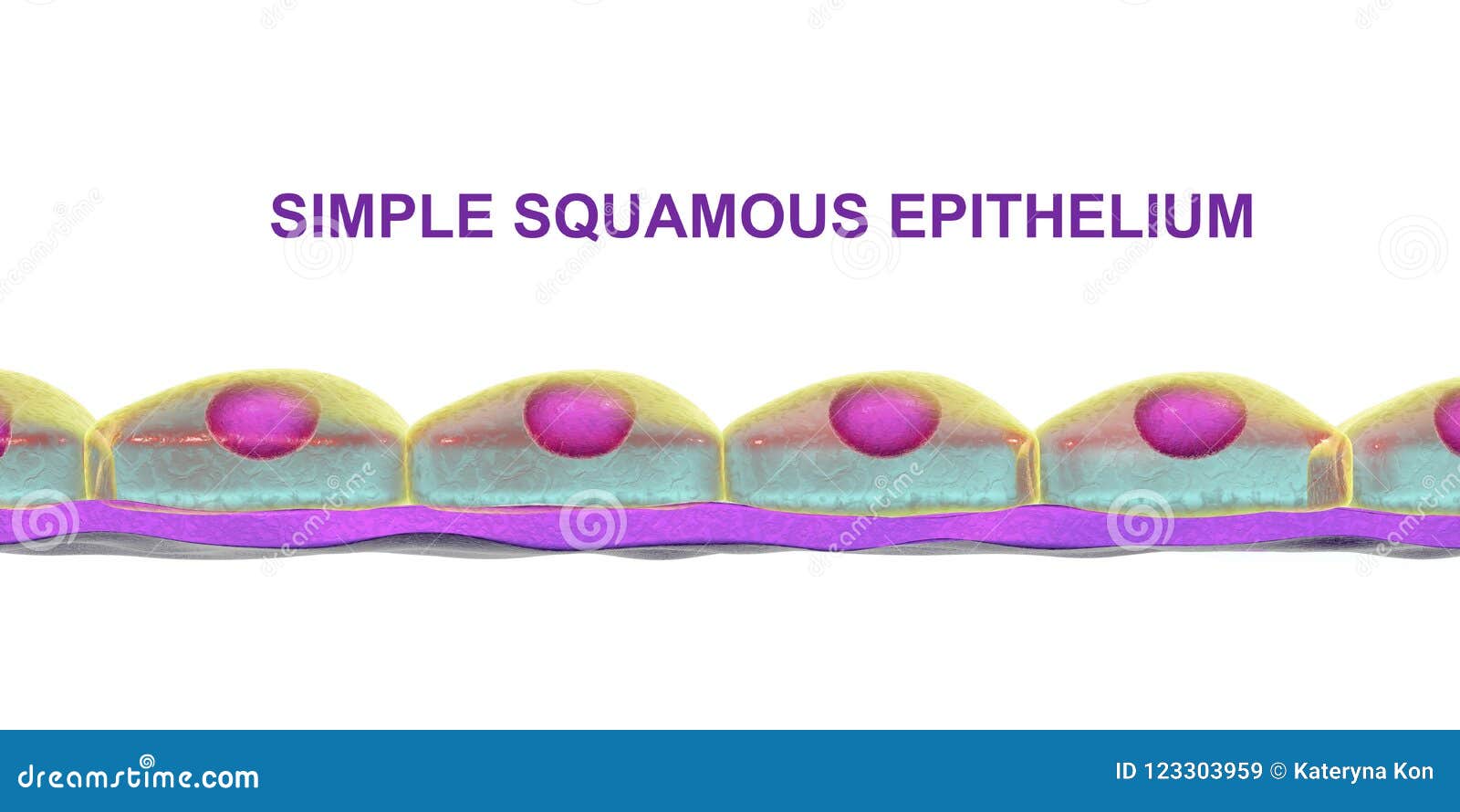
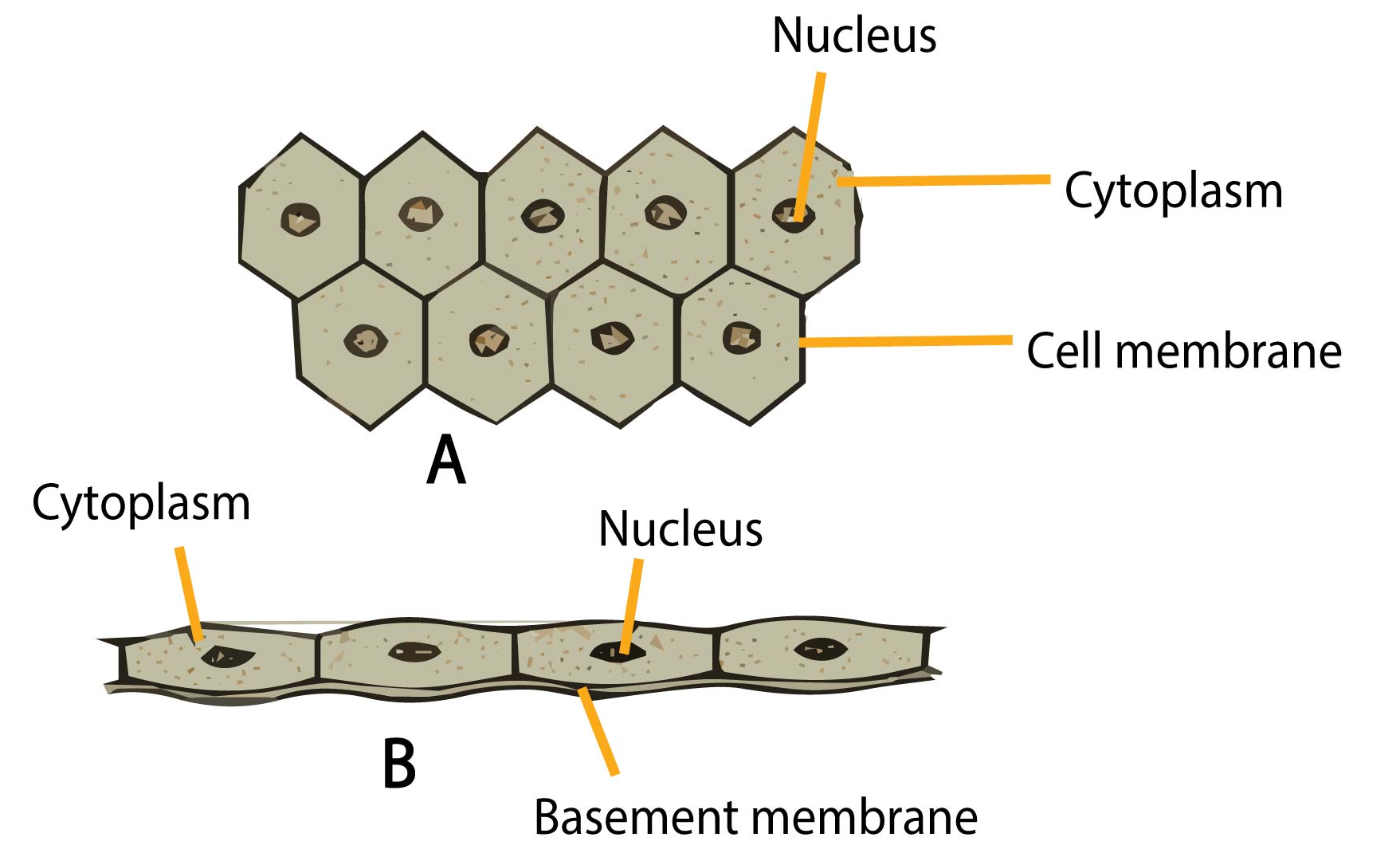
Simple squamous epithelium labeled diagram
The epithelium of the Thick segment is low simple cuboidal epithelium. The epithelium of the Thin segment is simple squamous. They can be distinguished from the vasa recta by the absence of blood, and they can be distinguished from the thick ascending limb by the thickness of the epithelium. Nomenclature
27.09.2020 · Simple squamous epithelium is a type of simple epithelium that is formed by a single layer of cells on a basement membrane. It is a type of epithelium formed by a single layer of squamous or flat cells present on a thin extracellular layer, called the basement membrane. This epithelium is also termed the pavement epithelium because the cells appear like tiles on …
09.11.2021 · Respiratory system (Systema respiratorum) The respiratory system, also called the pulmonary system, consists of several organs that function as a whole to oxygenate the body through the process of respiration (breathing).This process involves inhaling air and conducting it to the lungs where gas exchange occurs, in which oxygen is extracted from the air, and carbon …
Simple squamous epithelium labeled diagram.
These labelled diagrams should closely follow the current Science courses in histology, anatomy and ... squamous epithelium) ORIGIN: ectoderm lamina propria ... Covers the external surface. blood vessel lined by endothelium (simple squamous epithelium) ORIGIN: mesoderm lumen GENERALISED SECTION epithelium OF THE BODY connective tissue beneath ...
Microscope Simple Squamous Epithelium Labeled Diagram Written By MacPride Friday, December 25, 2020 Add Comment Edit. Epithelium Web Lab. What Are The Differences Of Simple And Stratified Tissue Sciencing. Epithelial Tissue Anatomy Physiology. Https Www Augusta Edu Scimath Biology Docs Animaltissues Pdf.
Stratified Squamous Diagram Examples of Simple Squamous Epithelium Kidney Slide Keratinized Labeled Labeled Lung Tissue Cells 400X Labeled Slide Lung Labeled Simple Squamous Epithelium Structure Anatomy Simple Squamous Epithelium Labeled Lung Tissue Black White Simple Squamous Epithelium Connective Tissue Apical View Cell Labeled
This article will discuss the histology of the simple epithelium. Contents. Simple squamous; Simple cuboidal; Simple columnar; Pseudostratified ...
Simple Squamous Epithelium Definition. Simple squamous epithelia are tissues formed from one layer of squamous cells that line surfaces. Squamous cells are large, thin, and flat and contain a rounded nucleus. Like other epithelial cells, they have polarity and contain a distinct apical surface with specialized membrane proteins.
The most variation is seen in the epithelium tissue layer of the mucosa. In the esophagus, the epithelium is stratified, squamous, and non-keratinizing, for protective purposes. In the stomach. the epithelium is simple columnar, and is organized into gastric pits …
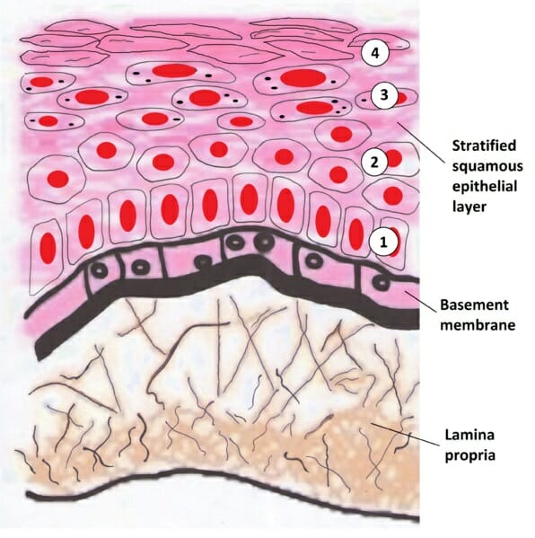
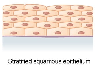
Squamous. stratified squamous diagram photo of endothelial cells. Squamous means scale-like. simple squamous epithelium is a single layer of flat ...
simple squamous epithelium diagram - Google Search. Find this Pin and more on Epithelial Tissue by Ileana Berezanski. Cool Car Backgrounds. Tissue Biology. Uk Board. Tissue Types. Cell Line. Photo Background Images. Body Tissues.
17.05.2021 · The simple squamous epithelium lines the surface of the lung tissue. You will find numerous thin-walled alveoli that line with the simple squamous epithelium. There are also intrapulmonary bronchus, three different types of bronchiole present with the lung tissue histology slide. You will find the mucosa, submucosa, hyaline cartilage layer, and adventitia in the …
Simple Squamous Epithelium. Function: Passage of materials by diffusion and filtration. Location: Air sacs of lungs. Simple Cuboidal Epithelium.7 pages
A simple squamous epithelium, also known as pavement epithelium, and tessellated epithelium is a single layer of flattened, polygonal cells in contact with ...
28.09.2020 · Plant Cell- Definition, Organelles, Structure, Parts, Functions, Labeled Diagram, Worksheet; Epithelial tissue vs Connective tissue- Definition, 15 Differences, Examples; Categories Biology Tags Epithelial Tissue, epithelium, squamous epithelium, Stratified epithelium Post navigation. Simple columnar epithelium- structure, functions, examples. …
05.10.2020 · The simple epithelial tissue is a closed network of flat epithelial cells. These are located on the basal membrane. It is composed of a single layer of cells that are specialized in diffusion, osmosis, filtration, secretion, and absorption.The simple epithelial tissue is found in the alveolar epithelium (pulmonary alveolus), the endothelium (lining of blood vessels and lymph …
Look at the areas outlined in the orientation diagram of the trachea and locate the loose, cellular connective tissue within the glands ... The area beneath the stratified squamous epithelium shown in slide 33 is the dermis, which is composed of dense irregular connective tissue. In this section, the fibers clearly predominate. This slide has been stained with iron hematoxylin and …
Simple Epithelium — The cells in simple squamous epithelium have the appearance of thin scales. ... This illustration shows a diagram of a goblet cell.
2 Sept 2021 — Figure 1 shows a diagram of simple squamous epithelium labeled. The tissue is polarized with one surface that faces the external environment ...
02.08.2015 · Draw a well labeled diagram to show various types of meristematic tissue and their location. 5. What type of tissue is found at the shoot apex?Name one more part of plant body where this type of tissue is found. 6. Why vacuoles are absent in the cells of meristematic tissue? 7. Do the roots of a plant continue to grow after their tips are removed?Give reason. …
Form the Outer Covering of the skin and some internal organs. Form the Inner Lining of blood vessels, ducts and body cavities, and the interior of the respiratory, digestive, urinary and reproductive systems. Glandular epithelia. Constitute the secretory portion of glands. Simple squamous epithelium. Most delicate epithelium.
Simple squamous epithelia consist of a single layer of flattened cells. This type of epithelia lines the inner surface of all blood vessels (endothelium), forms ...
Simple Squamous Epithelium (Figure 4.3a)A simple squamous epithelium is a single layer of flat cells. When viewed from above, the closely fitting cells resemble a tiled floor. When viewed in lateral section, they resemble fried eggs seen from the side.
















0 Response to "40 simple squamous epithelium labeled diagram"
Post a Comment