39 brain diagram reticular formation
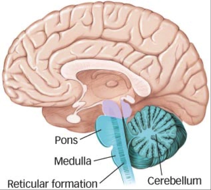
A net-like structure of mixed gray and white matter known as the reticular formation is found in all three regions of the brainstem. The reticular formation controls muscle tone in the body and acts as the switch between consciousness and sleep in the brain. The reticular (from the Latin reticulum, meaning net) formation is a far-reaching network of neurons extending from the spinal cord to the thalamus, with connections to the medulla oblongata, midbrain (mesencephalon), pons, and diencephalon.
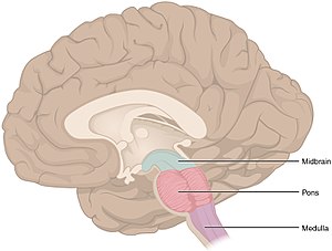
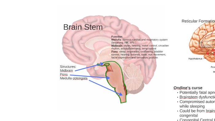
Reticular formation Cerebellum Hypothalamus Thalamus The Brainstem: •Medulla: controls breathing, heart rate, and blood pressure. •Pons: relays motor messages between cerebellum and motor cortex: influences sleep and dreaming. •Reticular Formation: affects arousal and attention. Cerebral Cortex THE BRAINSTEM
Brain diagram reticular formation
•* The reticular formation is a neural network located in ... Reticular formation Important Parts of the Brain Stem . The Midbrain/ Limbic System (The Mammalian Brain) •Located in the middle of Brain diagram labeled reticular formation. The reticular formation is a cluster of nerves within the brainstem that relay sensory and motor signals to and from the spinal cord and the brain. The skull consists of 22 bones 14 of which form the facial bones and the remaining. Activity in the brain stem is important for. Reticular Formation Helps control arousal, responds to change in monotony Thalamus Relays sensory information, switchboard between sensory neurons and higher brain regions Deals with sight, hearing, touch, taste. Transmits replies from higher brain to cerebellum and medullam Cerebellum
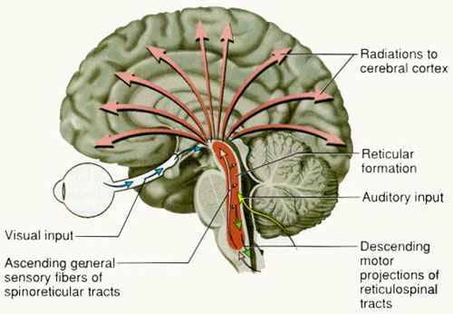
Brain diagram reticular formation. Ascending reticular activating system of the brain. Transl. Neurosci. Clin. 2016, 2(4): 275-285. 1 Reticular formation of the brain stem The difference between the electroencephalographic rhythms in awake and sleeping humans was initially described by Berger[1]. It became a breaking point in the development of sleep science or somnology. The the reticular formation, phylogenetically one of the oldest portions of the brain, is a poorly-differentiated area of the brain stem, centered roughly in the pons, but with the ascending reticular activating system connecting to areas in the thalamus, hypothalamus, and cortex, and the descending reticular activating system connecting to the … Labeled Diagrams of the Human Brain Central Core The central core consists of the thalamus, pons, cerebellum, reticular formation and medulla. These five regions are the central areas that regulate breathing, pulse, arousal, balance, sleep and early stages of processing sensory information. The reticular formation is made up of a net-like structure of various brainstem nuclei and neurons and covers an expansive portion of the brainstem, beginning in the mesencephalon, extending caudally through the medulla oblongata, and projecting into the superior cervical spinal cord segments.
The reticular formation is a cluster of nerves within the brainstem that relay sensory and motor signals to and from the spinal cord and the brain. This diagram labels the cranial nerves, including olfactory, oculomotor, trochlear, abducens . Describe the functions of the reticular formation region of the pons. Simple blank brain diagram. 85 of the brain is cerebral cortex divided as 41 frontal lobe 22 temporal lobe. Brain diagram with labels hypothalamus vector brain diagram pons cerebrum and cerebellum brain pons brain anatomy amygdala brain labelled amygdala brain human midbrain diagram pons. The brain is composed of 77 to 78 water and 10 to 12 lipids. The reticular formation is a cluster of nerves within the brainstem that relay sensory and motor signals to and from the spinal cord and the brain. It aids in the control of autonomic and endocrine functions, as well as muscle reflexes and sleep and awake states. The red nucleus is a mass of cells that aids in motor function. Substantia nigra ... The Reticular Formation. The reticular formation is a set of interconnected nuclei that are located throughout the brain stem. Its dorsal tegmental nuclei are in the midbrain while its central tegmental nuclei are in the pons and its central and inferior nuclei are found in the medulla. The Reticular Formation.
Brain Diagram(15 points) This is a hand drawn (in color) picture of the human brain that includes the following structures: medulla pons reticular formation cerebellum thalamus hypothalamus amygdala hippocampus primary somatosensory cortex primary motor cortex frontal lobe occipital lobe temporal lobe parietal lobe corpus callosum. The reticular formation is the medulla, pons and midbrain and comprises the brainstem core (also known as the tegmentum). It is an arrangement of neurons throughout he brain stem. The parvocellular neurons receive information relating to arousal Reticular Formation Diagram. angelo on December 3, 2021. Understanding The Brain A Work In Progress Professor Keith Kendrick Reticular Activating System Nervous System Parts System. Reticular Formation Reticular Formation Reticular Activating System Medicine. Diagram showing the lobes of the brain. Image Source: Wikimedia Commons. The parietal lobe is located at the top of the brain, between the frontal and occipital lobe. It consists of the somatosensory cortex and is responsible for integrating sensory information from different parts of the body, especially visual information related to navigation and spatial orientation.
Reticular Formation - The Definitive Guide | Biology Dictionary
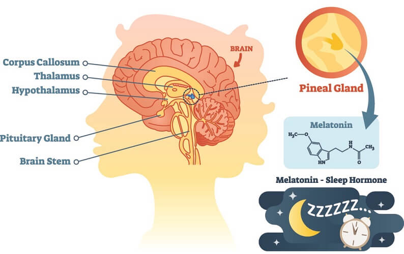
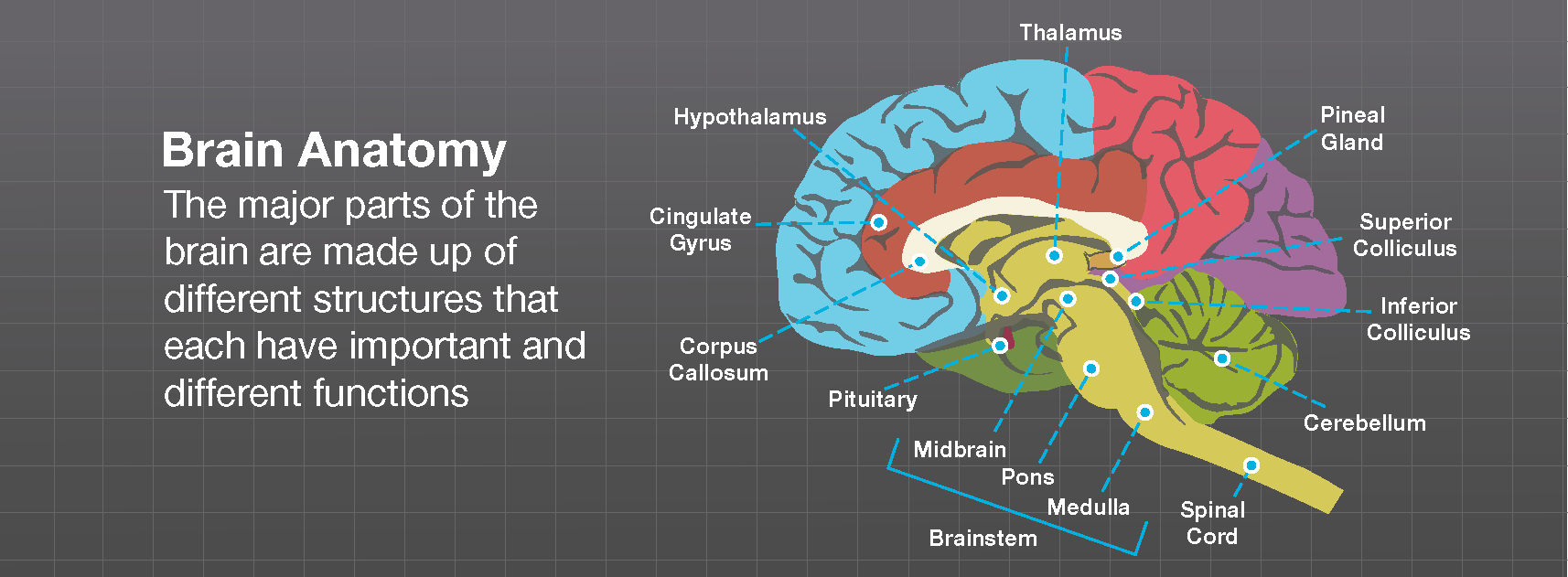
BRAIN PARTS - Notes from Textbook (Functions in chart/ Location on brain diagram) HINDBRAIN Medulla Mnemonics Pons Reticular Formation Cerebellum LIMBIC SYSTEM Thalamus Olfactory bulbs Hypothalamus Hippocampus Amygdala ("Ah-mig-duh-luh") Cingulate Cortex . CORTEX Corpus Callosum ...
Reticular Formation - The Definitive Guide | Biology Dictionary
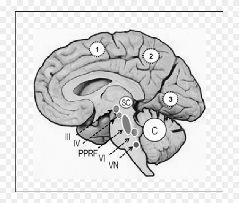
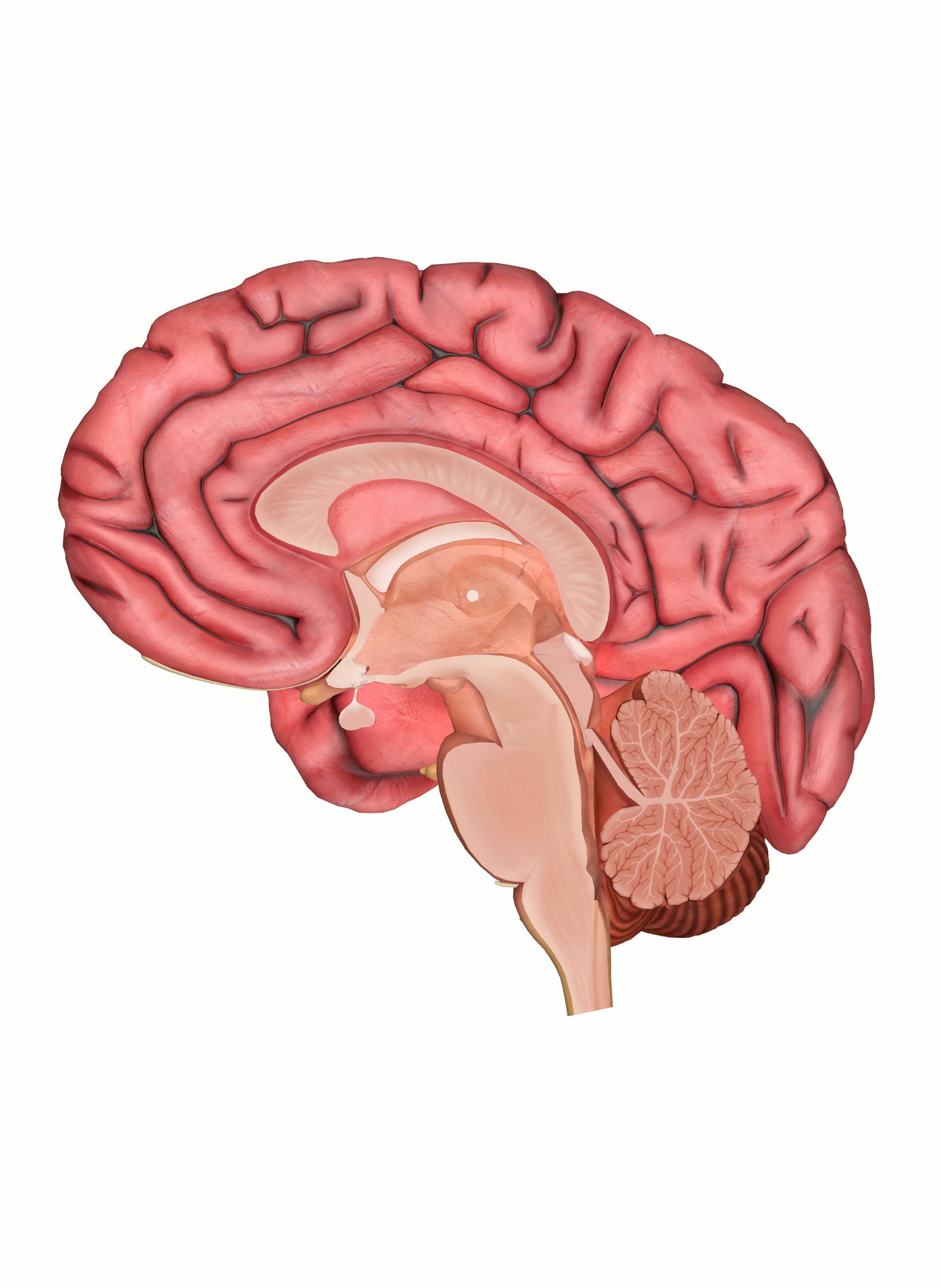
The reticular formation occupies a substantial amount of space in the dorsal half of the medulla. It is surrounded by the midline (medially), dorsomedial, dorsolateral, and lateral groups of nuclei and tracts mentioned above. The medial lemniscus occupies the ventral midline of the medulla.
Sagittal Section Of A Human Brain - Reticular Formation ...
The reticular formation is located in the brain stem. It extends throughout the length of the brainstem, along the central axis, from the spinal cord to the thalamus. It occupies the anterior portions of medulla, pons, midbrain, hypothalamus, and thalamus.

How the Brain Works Coherently, Under the Management of the ...
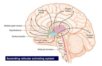
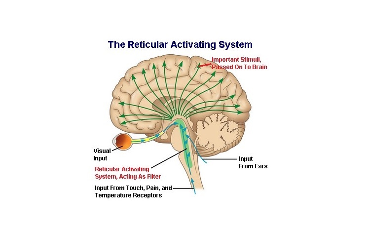
The Reticular Activating System (RAS) of the brain stem is considered as one of the most important systems which facilitates the functioning of sensation and attention. This is made up of a net-like bundle of neurons that run through the hind-brain, mid-brain and a part of the fore-brain called the hypothalamus.

Reticular Formation: Definition & Functions - Video & Lesson ...
Reticular formation: This highly diverse and integrative area contains a network of nuclei responsible for many vital functions including arousal, consciousness, sleep-wake cycles, coordination of certain movements, and cardiovascular control.; Periaqueductal gray (PAG) matter: This area plays a primary role in processing pain signals, autonomic function, and behavioral responses to fear and ...
ras [Brainstem Wiki]
The Brain Diagram Cerebrum Cerebellum Brain stem large part of your brain involving the four lobes "small brain" underneath the cerebrum in charge of basic vital functions like breathing 15 Terms Arthur_Hays3 Brain Diagram Reticular Formation Cerebellum Hypothalamus Controls arousal "Little brain" located below and behind the cerebrum; controls…
How Visualizing Something Into Existence Works - Gravity Learning
Organization of the Brainstem and Reticular Formation Competencies: In the brain stem, distinguish the locations of basilar, tegmentum and tectum components. Diagram a cross section of midbrain and locations of major tracts and nuclei. Construct a suprasegmental integrative system in the brain stem.
Brain Anatomy - Brainlab.org
Brain diagram reticular formation. It is also the origin of the descending analgesic pathways. The reticular formation cranial extension is upto the dienceph-alon subthalamus hypothalamus and. All the functions are carried out without a single glitch and before you. Peer Reviewed Journal High visibility Open Access.

Structure and Functions of the Reticular Activating System ...
The Reticular Formation Descending Reticular Formation Sleep and ArousalNeuronal Basis of Changes in the EEG Sleep Disorders The Ascending Reticular Activating System The brainstem contains many small neural networks that regulate essential functions, including the arousal system, cardiovascular and respiratory control, and the control of ...

Contributions to the Understanding of the Neural Bases of the ...
Our brain can be broken down into a variety of structures, the oldest of which is made up of the brainstem, thalamus, and the cerebellum.. The brainstem gives us our most basic functions like consciousness💭 (reticular formation), breathing and heartbeat ️ (medulla), and coordination of movement🏃 (pons).

Reticular Formation | Anatomy, Location, Structure & Physiology
The reticular formation is a set of interconnected nuclei that are located throughout the brainstem. It is not anatomically well defined, because it includes neurons located in different parts of the brain.
Motivation and emotion/Book/2014/Reticular formation, arousal ...
Here are a number of highest rated Reticular Formation Brain pictures on internet. We identified it from reliable source. Its submitted by organization in the best field. We admit this kind of Reticular Formation Brain graphic could possibly be the most trending subject as soon as we ration it in google improvement or facebook.

Anatomy of the Brain - NeuroICU RNs E-Learning/Resources
Reticular Formation Helps control arousal, responds to change in monotony Thalamus Relays sensory information, switchboard between sensory neurons and higher brain regions Deals with sight, hearing, touch, taste. Transmits replies from higher brain to cerebellum and medullam Cerebellum
Hindbrain | Brain for ai Wiki | Fandom
Brain diagram labeled reticular formation. The reticular formation is a cluster of nerves within the brainstem that relay sensory and motor signals to and from the spinal cord and the brain. The skull consists of 22 bones 14 of which form the facial bones and the remaining. Activity in the brain stem is important for.
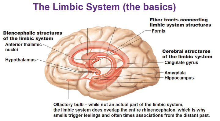
The Limbic System and the Reticular Formation
•* The reticular formation is a neural network located in ... Reticular formation Important Parts of the Brain Stem . The Midbrain/ Limbic System (The Mammalian Brain) •Located in the middle of

Brain anatomy, illustration - Stock Image - C046/1392 ...

Brain: Ultimate Guide to the Brain for AP® Psychology

FORMATIO RETICULARIS, RETICULAR FORMATION - ppt download

Reticular Formation: Definition & Functions - Video & Lesson ...
Brain – Human Brain Diagrams and Detailed Information

Reticular Activating System: Definition & Function Video

Reticular formation - human anatomy organs
Brain Stem & RAS by Andy Lim

Figure 3 from Reticular formation: A Review | Semantic Scholar

Ascending Reticular Activating System - an overview ...

Reticular formation and ascending reticular system (ARAS ...

Reticular Activation: How the Human Anatomy Prevents Ads from ...
Cns 14

Hindbrain: Parts, Function, and Location | Simply Psychology

reticular formation - Google zoeken | Reticular formation ...
Reticular formation: Anatomy and clinical notes | Kenhub
Reticular formation, Reticular activating system & Types of ...

Brain Anatomy - Internal Structures - Medical Art Library
The Brain, Cranial Nerves, and Sensory and Motor Pathways
THE RETICULAR FORMATION
What is reticular activating system (RAS)? - Quora

Brainstem, Thalamus, Reticular Formation & Cerebellum- The ...
reticular formation
Structures of the Brain Notes (Chapter 3)
0 Response to "39 brain diagram reticular formation"
Post a Comment