42 diagram of synovial joint
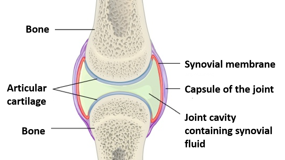
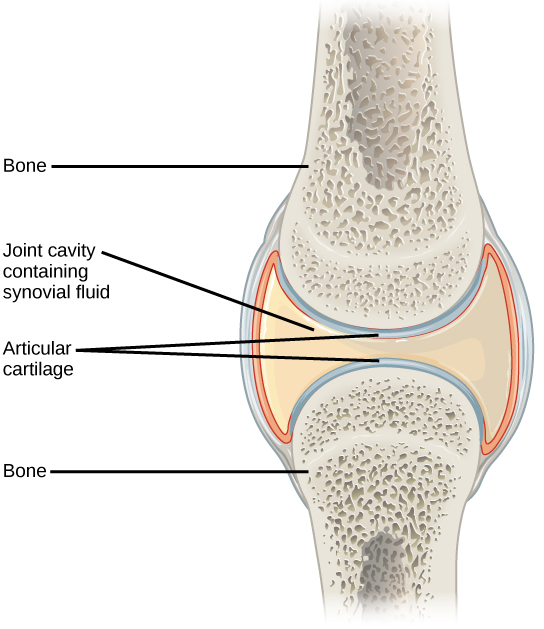
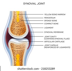
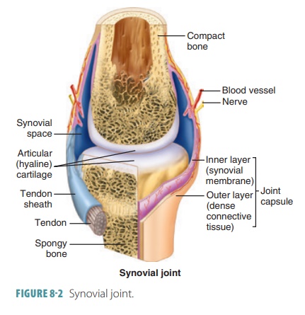
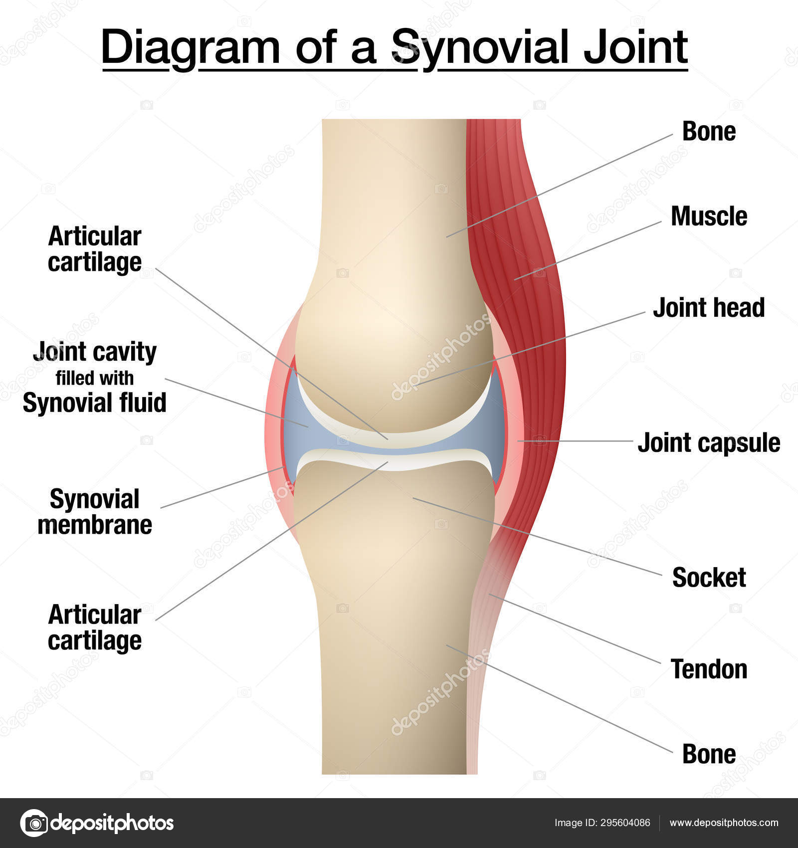
A synovial joint contains a synovial cavity and dense, irregular connective tissue that forms the articular capsule normally associated with accessory ligaments. Key Terms. articulation: A joint or the collection of joints at which something is articulated, or hinged, for bending. At synovial joints, the articular surfaces of bones are covered with smooth articular cartilage. This gives the bones of a synovial joint the ability to move smoothly against each other, allowing for increased joint mobility. Figure 9.4.1 – Synovial Joints: Synovial joints allow for smooth movements between the adjacent bones. The joint is surrounded by an articular capsule that defines a joint cavity filled with synovial fluid.
Synovial joint diagram Here in the diagram, you will find all the structures of a synovial joints in animals. If you need more diagram like this synovial joint then, you may follow anatomy learner blog or social media. Again, you might read other different article related to veterinary osteology or syndesmology with the anatomy learner.

Diagram of synovial joint
Synovial Joints. Joints can be simply defined as articulations of bones, which functions by providing shape to the skeleton system, protects bones by holding them together securely and also helps in movement. Based on structure and functions, joints have been further classified into different types. A synovial joint is one among the three types ... A synovial joint is a connection between two bones consisting of a cartilage lined cavity filled with fluid, which is known as a diarthrosis joint. Diarthrosis joints are the most flexible type of joint between bones, because the bones are not physically connected and can move more freely in relation to each other. The main parts of synovial joints are labelled on the synovial joint diagram and described in the table below. A, The egg-shaped ovoid surface represents a characteristic of most synovial joints of the body (e.g., hip joint, radiocarpal joint, knee joint, metacarpophalangeal joint). The diagram shows only the convex member of the joint.
Diagram of synovial joint. Nov 08, 2019 · Hinge joints allow bones to move in one direction back and forth, much like the hinge on a door. This article looks at their anatomy and function and includes an … Jan 01, 2019 · The bones of the hip include the femur, the ilium, the ischium, and the pubis. The pubis, ischium, and ilium together constitute the pelvis while the thigh bone is the femur. The bones together make up the hip. The hip itself is a ball and socket joint, much like the shoulder.The structures necessary to create this joint are the socket, the joint capsule, … ^ the joint capsule and synovial membrane become inflamed and articular cartilage is damaged or destroyed. Recommended textbook explanations. Introduction to Anatomy and Physiology Michelle Provost-Craig, Susan J. Hall, William C. Rose. 1,678 explanations. Anatomy and Physiology. The talocrural joint is a synovial hinge joint that connects the distal ends of the tibia and fibula in the lower limb with the proximal end of the talus. The articulation between the tibia and the talus bears more weight than that between the smaller fibula and the talus.
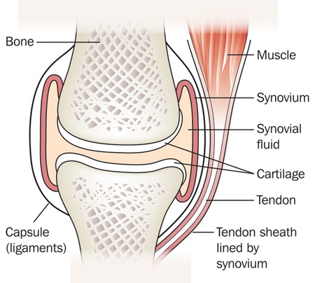
A synovial joint is characterised by the presence of a fluid-filled joint cavity contained within a fibrous capsule. It is the most common type of joint found in the human body, and contains several structures which are not seen in fibrous or cartilaginous joints.. In this article we shall look at the anatomy of a synovial joint – the joint capsule, neurovascular structures and clinical ... A synovial joint is the type of joint found between bones that move against each other, such as the joints of the limbs (e.g. shoulder, hip, elbow and knee). Characteristically it has a joint cavity filled with fluid. Other types of joint allow little or no movement, including fibrous joints (e.g between the bones of the skull) and ... Synovial Joint Definition. A synovial joint is a connection between two bones consisting of a cartilage lined cavity filled with fluid, which is known as a diarthrosis joint. Diarthrosis joints are the most flexible type of joint between bones, because the bones are not physically connected and can move more freely in relation to each other. Mar 20, 2021 · A normal knee joint is surrounded by a membrane, the synovium, which produces a small amount of thick fluid, known as synovial fluid. Synovial fluid helps to nourish the cartilage and keep it slippery. The synovium also has a tough outer layer (the joint capsule) which protects and supports the joint.
A synovial joint is also called diarthrosis, joint cartilage or bones with a fibrous joint. These joints allow bones to rotate around each other and slide past each other. The synovial joint has a joint cavity filled with fluid, together with muscles, ligament, tendons, and the capsule which keeps the bones of the joint in place. The synovial joint is a moveable or true joint in an animal's body. Hi there, do you want to learn synovial joint anatomy in animals? Fine, in this article, I will describe the synovial joint structure with a labeled diagram. I will also describe different types of synovial joints in animals. After reading this article, you will know the ... Solution. When the two bones are joined with the help of connective tissues which allows the movement of the bones is known as the synovial joint. This is a joint which is present between the long bones. The two bones are held with the help of fibrous tissue of the ligament. There is a presence of articular cartilage at the end of the long bones. 4. Which structure in the synovial joint; Question: 6. all are freely movable or diarthrotic 2. Label the diagram of a typical synovial joint using the terms provided in the key and the Key: a. articular capsule b. articular cartilage C. fibrous layer d. joint cavity e. ligament f. periosteum g. synovial membrane 3.

Synovial joints - human anatomy organs
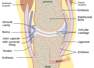
Apr 23, 2015 · The knee joint capsule is the structure surrounding the knee, made up of ligaments, bone, and fluid-filled cavities. It allows the full knee to have flexion, or bending motion, due to the folds ...

Draw labelled diagram Synovial joint. - Biology | Shaalaa.com
Classification of Joints • 1. According to the type of tissue at the joint: • a) Fibrous joint -- uses fibrous connective tissue to articulate bones. • b) Cartilaginous joint-- uses hyaline cartilage and/or fibro- cartilage to articulate bones. • c) Synovial joint --uses auricular cartilage, synovial membrane, joint capsule, and ligaments to articulate bones.

Synovial joint structure stock vector. Illustration of ...
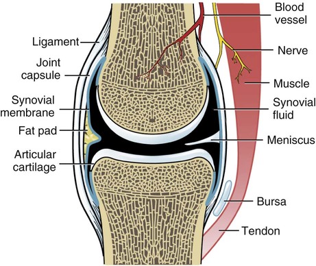
joints are characterized by the presence of a joint cavity (Figure 9). The cavity contains synovial fluid, enclosed within a tough fibrous capsule. ... View in ...

Synovial fluid - Wikipedia
Labelled Diagram Of Synovial Joint. A synovial joint is a connection between two bones consisting of a cartilage lined As seen in the above picture, the most powerful bite in the world gets its. A synovial joint or diarthrosis occurs at articulating bones to allow movement. fibrous connective tissue found in various parts of the body such as ...

Structures of a Synovial Joint - Capsule - Ligaments ...
The structure of a synovial joint is demonstrated by a diagram in which the articulating bones are surrounded by the articular capsule, which comprises an exterior fibrous capsule and an interior synovial membrane. Start studying label the synovial joint. Learn vocabulary, terms, and more with flashcards, games, and other study tools. ...

The structure of a synovial joint. | Synovial joint, Joints ...
Next, let's focus on hinge joints, shown as letter B on the diagram. Hinge joints are the synovial joint type referred to in our introductory section. These joints can be found between your upper ...

Synovial joint - labeled. | CanStock
Next, let's focus on hinge joints, shown as letter B on the diagram. Hinge joints are the synovial joint type referred to in our introductory section. These joints can be found between your upper and lower arm bones, otherwise called your elbow, as well as your ankles, fingers, toes, and knees. Hinge joints operate just like the hinges on a door.
Types of joints: Anatomy and arthrology | Kenhub
Synovial joints. A joint is a place where two or more bones meet and is also called an articulation. The role of joints and connective tissue . Connective tissues consist of ligaments, cartilage ...

Synovial Joint: structure and label Diagram | Quizlet
The only 2 synovial joints that aren't diarthrotic. carpals and tarsals. Functional classification of carpals and tarsals. amphiarthrosis. membrane continuous from bone to bone outside the articular capsule. periosteum. fibrous capsule lined by synovial membrane. articular capsule.

Synovial Joint Mechanics | Musculoskeletal Key
Aug 14, 2020 · The bones of a synovial joint are surrounded by a synovial capsule, which secretes synovial fluid to lubricate and nourish the joint while acting as a shock absorber. The ends of the joint bones are covered with smooth, glass-like hyaline cartilage which reduces friction during movement. A synovial joint contains a synovial cavity and dense, irregular connective tissue that forms the articular capsule normally associated with accessory ligaments.

Sketch and label typical synovial joint. - Brainly.in
Download Synovial Joint Diagram Labeled Cartoon Vector via CartoonDealer. Educational Otherwise Tool Wonderfully Presented Designed. Zoom into our collection of high-resolution cartoons, stock photos and vector illustrations. Image: 39898469

Chapter 8.4.1 General Structure of Synovial Joints BIO201
Sep 18, 2018 · Labelled Diagram Of Synovial Joint. The structure and function of synovial joints is our second dash point under the skeletal system. The skeletal system has a number of different. A synovial joint is a connection between two bones consisting of a cartilage lined As seen in the above picture, the most powerful bite in the world gets its. A synovial joint is a connection between two bones consisting of a cartilage lined As seen in the above picture, the most powerful bite in the world gets its.
Joints
Synovial Joint. Create healthcare diagrams like this example called Synovial Joint in minutes with SmartDraw. SmartDraw includes 1000s of professional healthcare and anatomy chart templates that you can modify and make your own. 26/37 EXAMPLES.

Synovial Joints: Structure, Function & Types | Study.com
The given diagram of the knee joint can help you to understand its various parts and the description given below will give you an insight of the functioning of the knee. ⚫ Bone There are three bones in the knee namely the femur which is the thigh bone, tibia which is the shin bone and patella which is the knee cap.

Synovial joint anatomy stock vector. Illustration of science ...
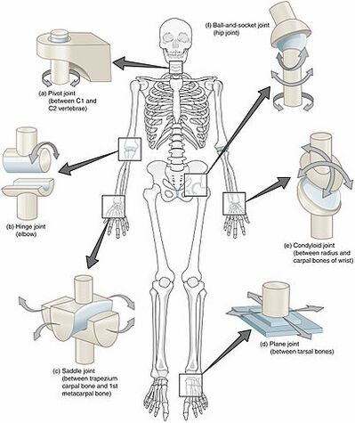
Synovial joints are often further classified by the type of movements they permit. There are six such classifications: hinge (elbow), saddle (carpometacarpal ...
Joint: synovial - MyDr.com.au
A joint or articulation (or articular surface) is the connection made between bones in the body which link the skeletal system into a functional whole. They are constructed to allow for different degrees and types of movement. Some joints, such as the knee, elbow, and shoulder, are self-lubricating, almost frictionless, and are able to withstand compression and maintain heavy …

Osteoarthritis Healthy Synovial Joint And Knee With Arthritis ...
Jan 02, 2022 · The synovial joint is a moveable or true joint in an animals body. Key Structures of a Synovial Joint. Produced by the synovial membrane to lubricate the joint. Labelled Diagram Of Synovial Joint. Define and describe dynamic movements that occur at synovial joints. Draw a labelled diagram of a synovial joint. Anatomy of a synovial joint.

The Development of Synovial Joints - ScienceDirect
The main parts of synovial joints are labelled on the synovial joint diagram and described in the table below. A, The egg-shaped ovoid surface represents a characteristic of most synovial joints of the body (e.g., hip joint, radiocarpal joint, knee joint, metacarpophalangeal joint). The diagram shows only the convex member of the joint.

Figure 8.3 General structure of a synovial joint.
A synovial joint is a connection between two bones consisting of a cartilage lined cavity filled with fluid, which is known as a diarthrosis joint. Diarthrosis joints are the most flexible type of joint between bones, because the bones are not physically connected and can move more freely in relation to each other.
![A typical synovial joint (the knee joint) [19]. | Download ...](https://www.researchgate.net/profile/Houssein-Lamine-2/publication/339686957/figure/fig4/AS:865361939402752@1583329772113/A-typical-synovial-joint-the-knee-joint-19.jpg)
A typical synovial joint (the knee joint) [19]. | Download ...
Synovial Joints. Joints can be simply defined as articulations of bones, which functions by providing shape to the skeleton system, protects bones by holding them together securely and also helps in movement. Based on structure and functions, joints have been further classified into different types. A synovial joint is one among the three types ...
Synovial Joint - Types - Characteristic features - AnatomyQA
What type of acid is in synovial fluid? + Example

Joints | BioNinja
19.3 Joints and Skeletal Movement – Concepts of Biology – 1st ...
Synovial joints Images, Stock Photos & Vectors | Shutterstock

A general synovial joint. | Download Scientific Diagram

Describe typical synovial joint with a neat labelled diagram ...

Anatomy: Synovial Joint Diagram Diagram | Quizlet

Types of Synovial Joints | Biology for Majors II
Joint - Wikipedia
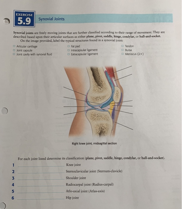
Solved EXERCISE Synovial Joints Synovial joints are freely ...
Joints
Structure and Function of Joints | Musculoskeletal Key

Figure 2.1 from Literature Review 2.1 Synovial Joints ...
Synovial Joints

Synovial fluid in joints: what it is and how exercise affects ...

Synovial joint - Wikipedia
Synovial Joints - Physiopedia

Synovial (movable) Joints
Synovial joint chart. Labeled anatomy infographic with two ...
Elbow synovial joint - Labelled diagram
Chapter 8
0 Response to "42 diagram of synovial joint"
Post a Comment