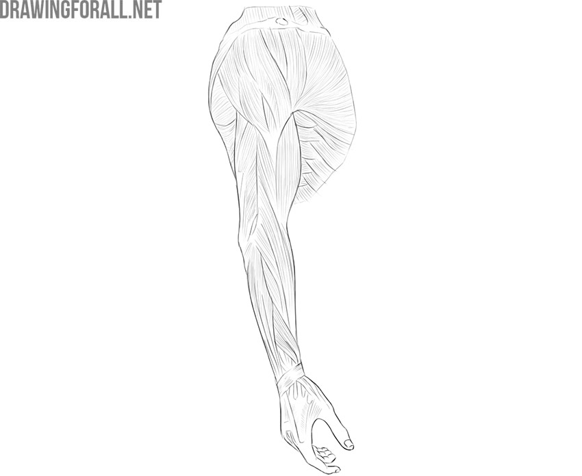
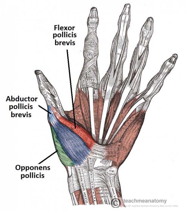
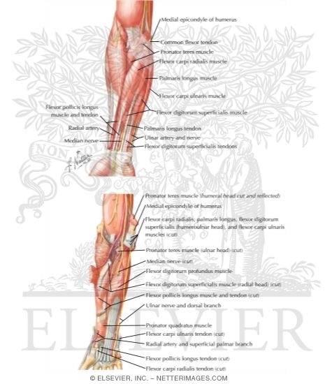
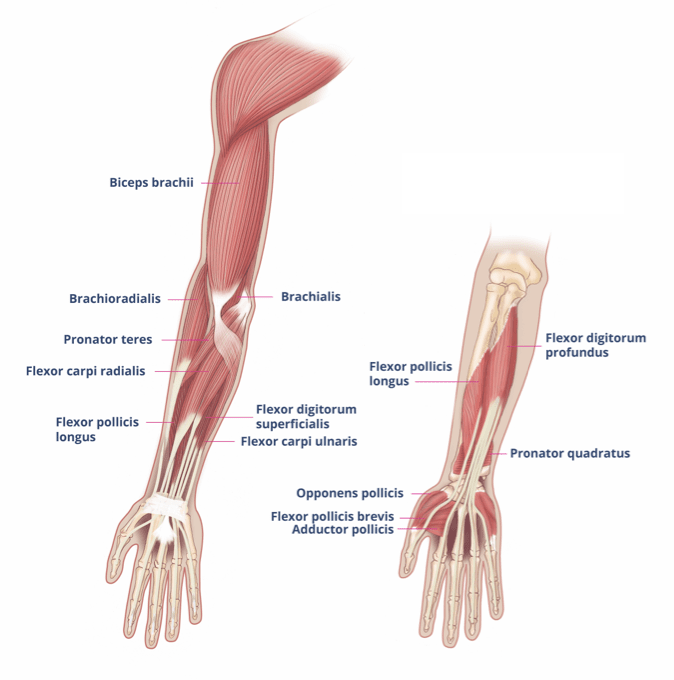
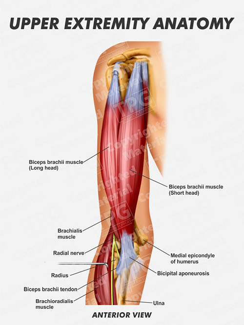
42 Muscles Of Upper Limb Diagram
Leg Muscles Anatomy, Function & Diagram | Body Maps The majority of muscles in the leg are considered long muscles, in that they stretch great distances. As these muscles contract and relax, they move skeletal bones to create movement of the body. Dissection of Rat (With Diagram) | Zoology ADVERTISEMENTS: In this article we will discuss about the dissection of rat. Also learn about:- 1. Dissection of Alimentary System 2. Dissection of Circulatory System 3. Dissection of Venous System 4. The Arterial System 5. Dissection of Cranial Nerves 6. Dissection of Brain 7. Dissection of Neck Region 8. Dissection of Urinogenital System 9. The […]
› en › libraryLearn the muscles of the arm with quizzes & diagrams | Kenhub Jan 24, 2022 · In today’s case, our tool of choice is a diagram with the arm muscles clearly labeled. Seeing all of the muscles together in one diagram is a great way to train your brain to differentiate between all of the different structures so you don’t get them jumbled up later down the line.

Muscles of upper limb diagram
PDF Clinical Anatomy of the Upper Limb - Welcome to KHIMA •Clinical Anatomy of Nerve affect Upper Limb Muscles •Special Diagnostic Tests. Bones and Joints of Upper Limb Regions Bones Joints Shoulder Girdle Clavicle Scapula Sternoclavicular Joint Acromioclavicular Joint Bones of Arm Humerus Upper End: Glenohumeral Joint Lower End: See below Bones of Forearm Radius Dog Leg Anatomy with Labeled Diagram - Bones, Joints ... 7.9.2021 · Dog leg anatomy. First, you might have a basic idea of the different bones of the forelimb and hindlimb of a dog. Now I will provide you the few information on the other bones of dog leg anatomy with their unique features. The front leg of a dog consists of the clavicle, scapula (arm), radius and ulna (forearm), carpals, metacarpals, and phalanges (forepaw). › science › human-muscle-systemhuman muscle system | Functions, Diagram, & Facts | Britannica Human muscle system, the muscles of the human body that work the skeletal system, that are under voluntary control, and that are concerned with movement, posture, and balance. Broadly considered, human muscle—like the muscles of all vertebrates—is often divided into striated muscle, smooth muscle, and cardiac muscle.
Muscles of upper limb diagram. Bones of the Hand - Carpals - Metacarpals - TeachMeAnatomy 16.8.2020 · Collectively, the carpal bones form an arch in the coronal plane. A membranous band, the flexor retinaculum, spans between the medial and lateral edges of the arch, forming the carpal tunnel.. Proximally, the scaphoid and lunate articulate with the radius to form the wrist joint (also known as the ‘radio-carpal joint’). In the distal row, all of the carpal bones articulate with the ... Anatomy Tables - Muscles of the Upper Limb a respiratory muscle, it receives ventral ramus innervation; embryonically related to the intercostal muscles, not the deep back mm. serratus posterior superior : ligamentum nuchae, spines of vertebrae C7 and T1-T3 : ribs 1-4, lateral to the angles: elevates the upper ribs: branches of the ventral primary rami of spinal nerves T1-T4 UPPER LIMB - Yavapai College Diagram and follow the path of spinal nerves through the brachial plexus out to the muscles of the upper limb; Diagram the path of sensory innervation from regions of the upper limb through the brachial plexus; Predict the type of condition/impairment caused by nerve damage to main branches of brachial plexus ... Muscles of the larynx: Anatomy, function, diagram | Kenhub 14.2.2022 · Muscles of the larynx. There are many muscles that either make up a certain part of the laryngeal structure inside the neck, or that sit adjacent to it and aid in its function.These muscles produce the movements of the larynx and its cartilages, thus enabling the proper air conduction, speech, movements of the epiglottis and airways protection.
Skeleton of upper limb | Encyclopedia | Anatomy.app ... Free upper extremity. The upper aspect of the free upper extremity is formed by the upper arm that consists of a single bone called the humerus. The middle part of the free upper extremity is known as the forearm, and it is made of two parallel lying bones - radius and ulna. Human Upper Body Muscle Diagram - Studying Diagrams New users enjoy 60 OFF. We hope this picture Human upper extremity muscle diagram can help you study and research. Lower Body Muscles Diagram Jpg 500 630 Anatomiya Cheloveka. Human muscle system the muscles of the human body that work the skeletal system that are under voluntary control and that are concerned with movement posture and balance. PDF Reference Diagrams of Upper Limb Muscles: Names, Locations ... Page 18 A25LAB EXERCISES: UPPER LIMB MUSCLES A25LAB EXERCISES: UPPER LIMB MUSCLES Page 19 LAB ASSIGNMENT 3: UPPER LIMB MUSCLE IDENTIFICATION (MODELS) The following diagrams are based on the intact upper limb models (Somso). Color each muscle in the diagram, write its name on the diagram, then find it on the model. Muscles of Upper Extremity (Posterior Deep view) Muscles of Upper Extremity (Posterior Deep view) Click on a muscle's name to see detailed information or Take a Quiz.
Anatomy, Shoulder and Upper Limb, Muscles - StatPearls ... The upper limb comprises many muscles which are organized into anatomical compartments. These muscles act on the various joints of the hand, arm, and shoulder, maintaining tone, providing stability and allowing precise fluid movement. Axioappendicular groups of muscles arise from the axial skeleton to act upon the pectoral girdle. › site › handlersMuscles - Cabarrus County Schools 1. There are over 1,000 muscles in your body. -False. There are over 600 muscles in the body. 2. Skeletal, or voluntary, muscles are the muscles you can control. True. You can control your skeletal muscles to walk, run, pick up things, play an instrument, throw a baseball, kick a soccer ball, push a lawnmower, or ride a bicycle 3. Muscles of the Hand - 3D Models, Video Tutorials & Notes ... In the intrinsic muscles of the hand, you've got the hypothenar muscles, the thenar muscles, lumbricals, interosseous muscles, the palmaris brevis and the adductor pollicis. I'll just talk you through those in this tutorial. We're just looking at a diagram of the left hand, anterior view. Skeletal Muscles of Upper Extremity Diagram | Quizlet Muscles of the Upper Extremity Learn with flashcards, games, and more — for free.
List of skeletal muscles of the human body - Wikipedia There are also vestigial muscles that are present in some people but absent in others, such as palmaris longus muscle. [4] [5] The muscles of the human body can be categorized into a number of groups which include muscles relating to the head and neck, muscles of the torso or trunk, muscles of the upper limbs, and muscles of the lower limbs.
Forces and Torques in Muscles and Joints | Physics Muscles, bones, and joints are some of the most interesting applications of statics. There are some surprises. Muscles, for example, exert far greater forces than we might think. Figure 1 shows a forearm holding a book and a schematic diagram of an analogous lever system.
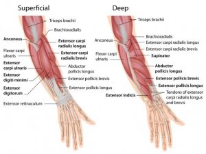
The Muscles of the Upper Limbs - Free Anatomy Quiz Quizzes on the muscles of the upper limb. The quizzes below each include 15 multiple-choice identification questions related to the muscles of the upper limbs, and includes the following muscles : The Abductor pollicis longus, Anconeus, Biceps brachii, Brachialis, Brachioradialis, Extensor carpi radialis brevis, Extensor carpi radialis longus ...
Muscles of the Upper Limb (Anterior View) Diagram | Quizlet Start studying Muscles of the Upper Limb (Anterior View). Learn vocabulary, terms, and more with flashcards, games, and other study tools.
Muscles of the Upper Arm - Anatomy Tutorial - YouTube 3D anatomy tutorial on the muscles of the upper arm using. Tutorial covers the muscles of the anterior and posterior compartment of the upper arm, and talks ...
teachmeanatomy.info › lower-limb › musclesMuscles of the Anterior Thigh - Quadriceps - TeachMeAnatomy Apr 20, 2020 · The muscles in the anterior compartment of the thigh are innervated by the femoral nerve (L2-L4), and as a general rule, act to extend the leg at the knee joint. There are three major muscles in the anterior thigh – the pectineus , sartorius and quadriceps femoris .
Smooth muscle: Structure, function, location | Kenhub 28.10.2021 · Smooth muscle (Textus muscularis levis) Smooth muscle is a type of tissue found in the walls of hollow organs, such as the intestines, uterus and stomach.. You can also find smooth muscle in the walls of passageways, including arteries and veins of de cardiovascular system.This type of involuntary non-striated muscle is also found in the tracts of the urinary, respiratory and …
Learn all muscles with quizzes and labeled diagrams | Kenhub If you're looking for a speedy way to learn muscle anatomy, look no further than our anatomy crash courses. Let's take a look at how you can use muscle diagrams for maximum benefit. Labeled diagram. View the muscles of the upper and lower extremity in the diagrams below. Use the location, shape and surrounding structures to help you memorize ...
Muscles of the Upper Arm - Biceps - Triceps - TeachMeAnatomy There are three muscles located in the anterior compartment of the upper arm - biceps brachii, coracobrachialis and brachialis. They are all innervated by the musculocutaneous nerve . A good memory aid for this is BBC - b iceps, b rachialis, c oracobrachialis.
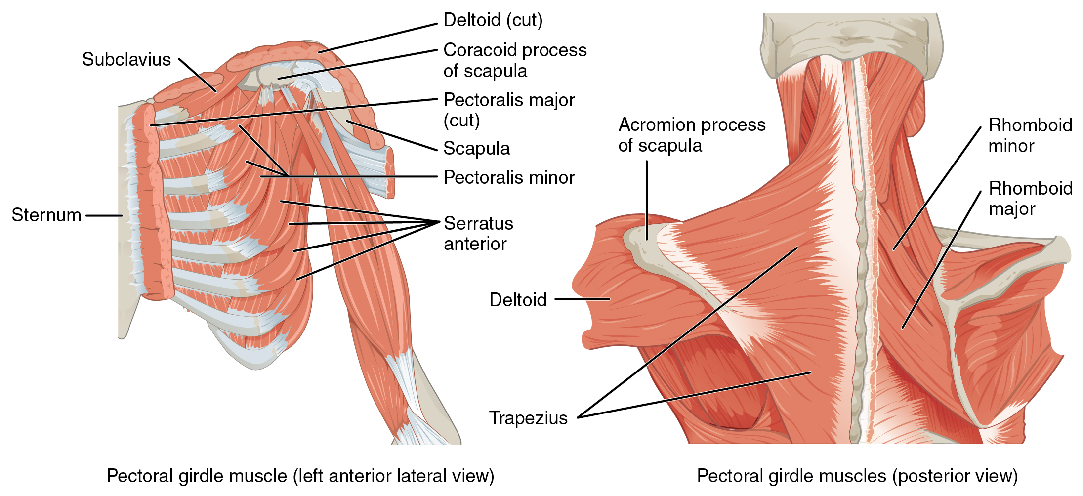
PDF Muscles Stabilizing Pectoral Girdle Muscles of the Upper Limb 11/8/2012 1 Muscles of the Upper Limb Pectoralis minor ORIGIN: anterior surface of ribs 3 - 5 ACTION INSERTION: coracoid process (scapula) Muscles Stabilizing Pectoral Girdle
Upper limb anatomy - eAnatomy - IMAIOS The study of the innervation of the upper limb begins with a diagram of the brachial plexus, then the paths of each nerve in the arm and the forearm are detailed (musculocutaneous nerve, median nerve, ulnar nerve, axillary nerve and radial nerve).
PDF The Muscles of the Upper Limbs - Free Anatomy Quiz The Muscles of the Upper Limbs Practice: Name the muscles in the diagrams below, using the list underneath. Choose from: Abductor pollicis longus Brachioradialis Extensor digiti minimi Flexor digitorum superficialis
› en › libraryMuscles of the leg quizzes and labeled diagrams | Kenhub Jan 24, 2022 · Leg muscles labeled. Take a look at the leg muscles diagram below, where you see each muscle clearly labeled. Spend some time revising this diagram by connecting the name and location of each structure with what you’ve just learned in the video. The aim of this exercise is to improve your confidence in identifying different structures.
11 Muscles of upper limb ideas | muscle anatomy, anatomy ... Nov 8, 2016 - Explore Alexandra Southard's board "Muscles of upper limb" on Pinterest. See more ideas about muscle anatomy, anatomy and physiology, massage therapy.
Upper limb anatomy: Bones, muscles and nerves | Kenhub The intrinsic muscles of the hand are the: palmaris brevis, interossei ( palmar and dorsal ), adductor pollicis, thenar, hypothenar and lumbrical muscles. You can learn everything about them with our learning materials and test yourself with the integrated quiz. Muscles of the hand Explore study unit
Muscles of Upper Extremity (Anterior Superficial view) Muscles of Upper Extremity (Anterior Superficial view) Click on a muscle's name to see detailed information or Take a Quiz.
› afp › 2010Peripheral Nerve Entrapment and Injury in the Upper Extremity ... Jan 15, 2010 · Peripheral nerve injury of the upper extremity commonly occurs in patients who participate in recreational (e.g., sports) and occupational activities. Nerve injury should be considered when a ...
PDF The Upper Limb Lecture 1 - anatomy.plcnet.org Arteries of upper limb Axillary artery Continuation of subclavian artery at lateral border of first rib Becomes brachial artery at lower border of teres major Divided into three parts by overlying pectoralis minor First portion, above muscle-gives rise to thoracoacromial a. Second portion, behind muscle-gives rise to lateral thoracic a.
Muscle Charts of the Human Body - PT Direct Muscle Charts of the Human Body For your reference value these charts show the major superficial and deep muscles of the human body. Superficial and deep anterior muscles of upper body
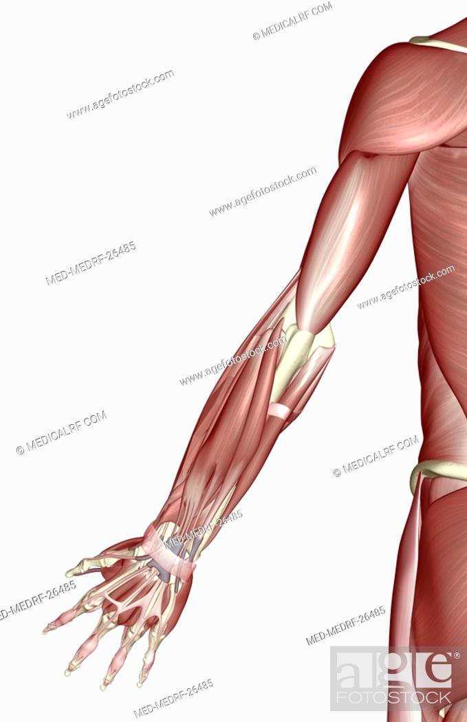
Muscles of Upper Limb Unlabeled | Muscles of upper limb ... Label the muscles of the arm. Designed for an anatomy class, two images of the arms can be colored and labeled, one focusing on the flexors and the other on the extensors. Anatomy charts are visual depictions of the human body. They can be used to illustrate an entire system or a specific body part or condition.
Muscles of the Upper Limb - TeachMeAnatomy The muscles of the upper limb can be divided into 6 different regions: pectoral, shoulder, upper arm, anterior forearm, posterior forearm, and the hand.. There are 4 muscles of the pectoral region: pectoralis major, pectoralis minor, serratus anterior and subclavius.Collectively, these muscles are involved in movement and stabilisation of the scapula, as well as movements of the upper limb.
Upper Limb - Important Questions , Anatomy QA Muscles forming musculotendinous cuff/rotator cuff and their nerve supply. Muscles of anterior/flexor compartment of arm and their nerve supply/ muscles supplied by musculocutaneous nerve. Muscle attached to the medial border of scapula. Structures attached to coracoid process of scap ula. Muscles responsible for abduction at shoulder joint.
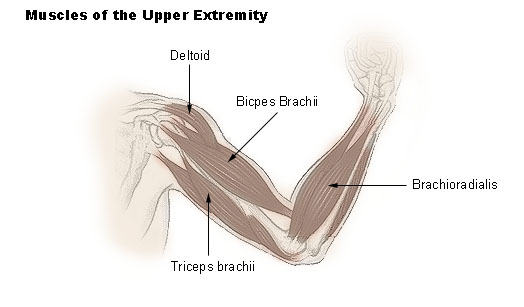
Muscles of the Upper Extremity | SEER Training The illustration below shows some of the muscles of the upper extremity. Muscles that move the shoulder and arm include the trapezius and serratus anterior. The pectoralis major, latissimus dorsi, deltoid, and rotator cuff muscles connect to the humerus and move the arm.
› science › human-muscle-systemhuman muscle system | Functions, Diagram, & Facts | Britannica Human muscle system, the muscles of the human body that work the skeletal system, that are under voluntary control, and that are concerned with movement, posture, and balance. Broadly considered, human muscle—like the muscles of all vertebrates—is often divided into striated muscle, smooth muscle, and cardiac muscle.
Dog Leg Anatomy with Labeled Diagram - Bones, Joints ... 7.9.2021 · Dog leg anatomy. First, you might have a basic idea of the different bones of the forelimb and hindlimb of a dog. Now I will provide you the few information on the other bones of dog leg anatomy with their unique features. The front leg of a dog consists of the clavicle, scapula (arm), radius and ulna (forearm), carpals, metacarpals, and phalanges (forepaw).
PDF Clinical Anatomy of the Upper Limb - Welcome to KHIMA •Clinical Anatomy of Nerve affect Upper Limb Muscles •Special Diagnostic Tests. Bones and Joints of Upper Limb Regions Bones Joints Shoulder Girdle Clavicle Scapula Sternoclavicular Joint Acromioclavicular Joint Bones of Arm Humerus Upper End: Glenohumeral Joint Lower End: See below Bones of Forearm Radius













0 Response to "42 Muscles Of Upper Limb Diagram"
Post a Comment