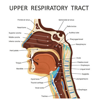
42 throat diagram front view
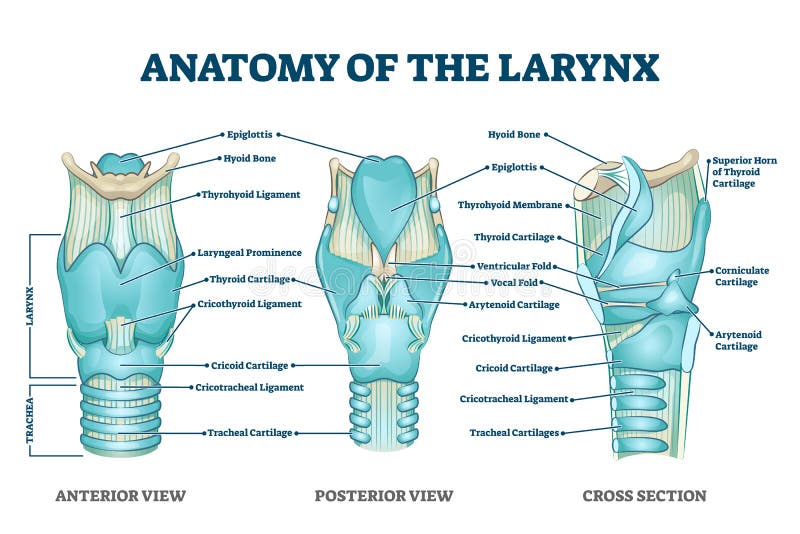
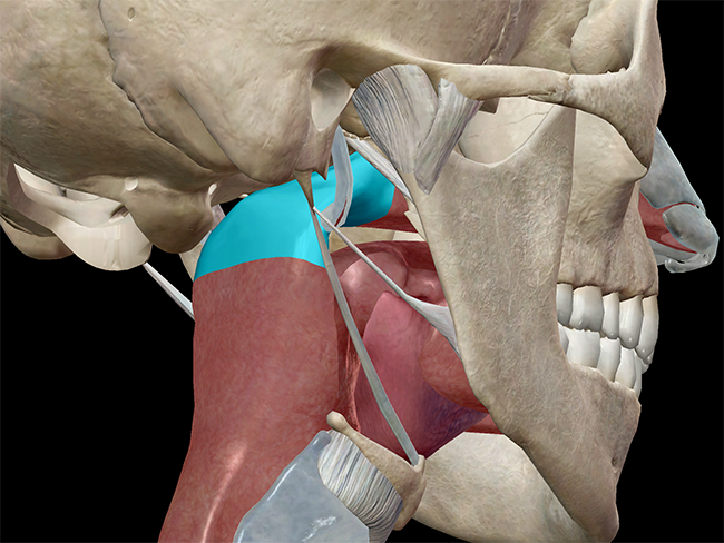
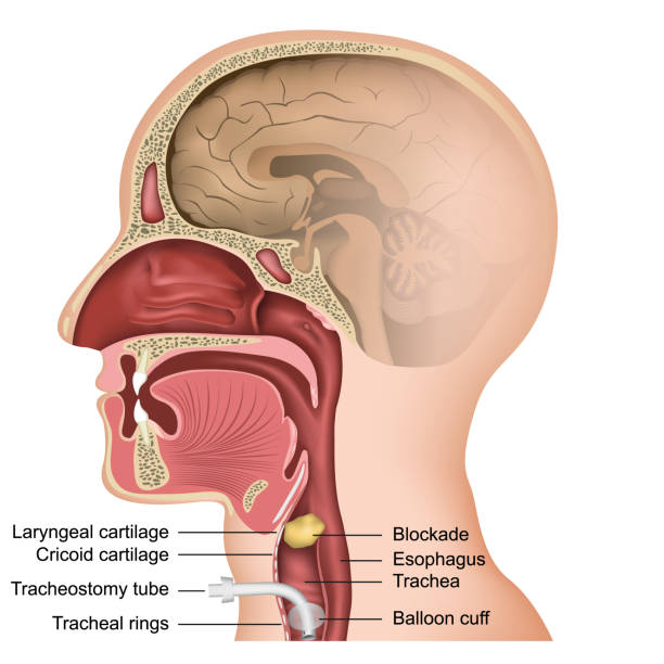
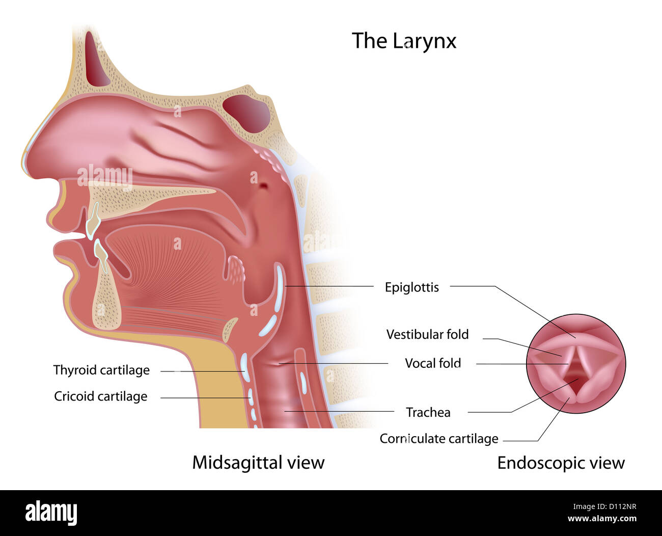
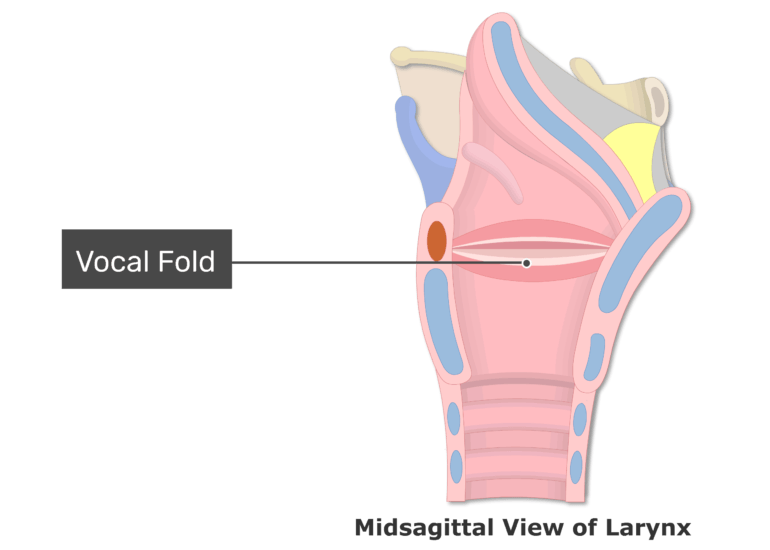
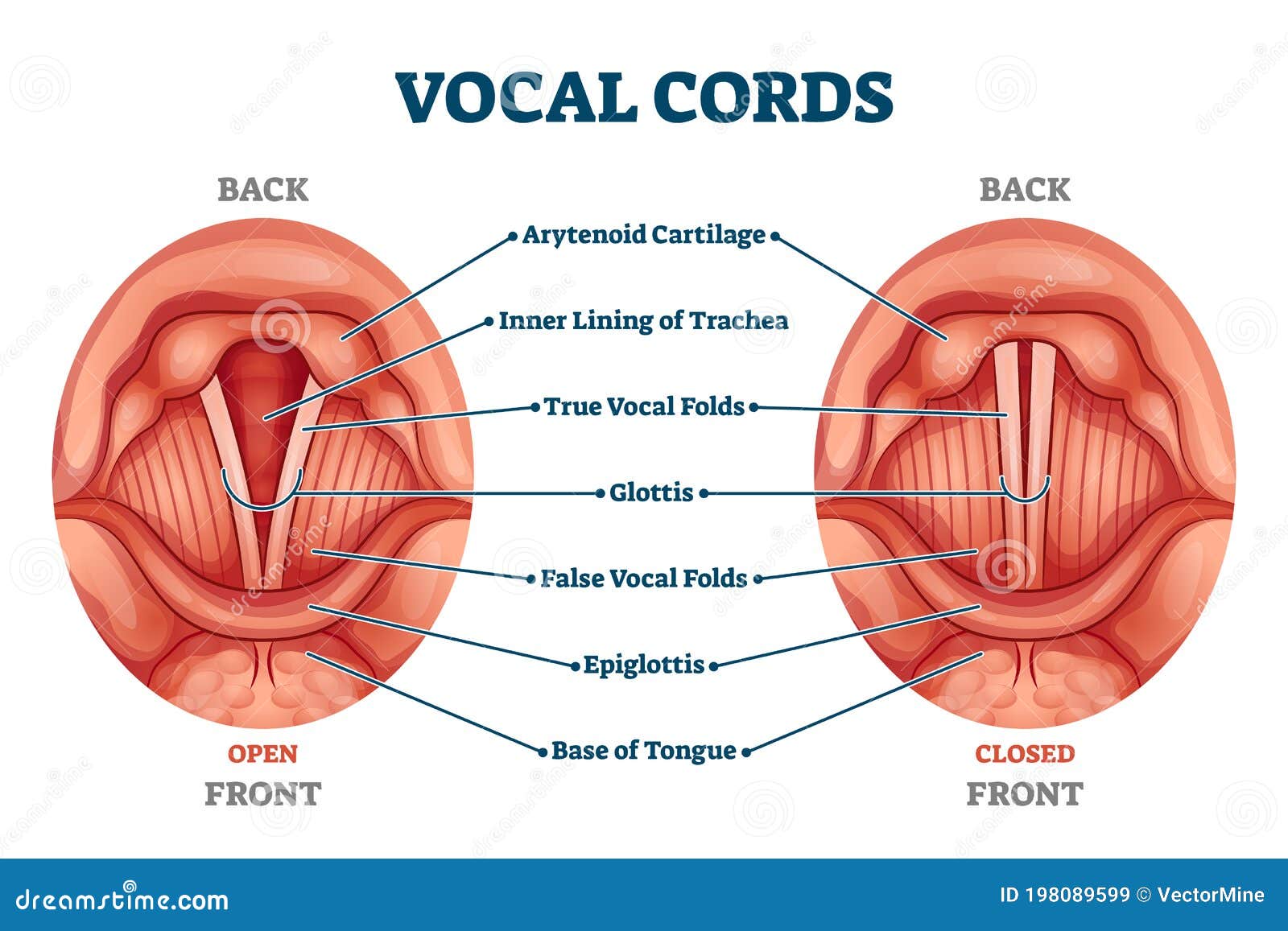
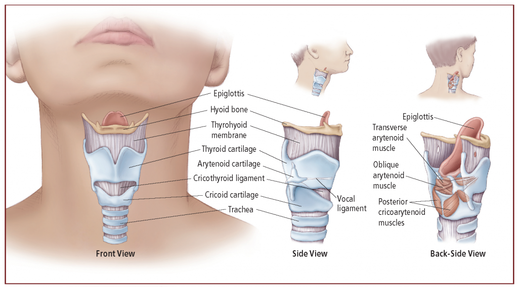
The Hyoid Bone: Anatomy, Function, and Conditions The hyoid bone is a small horseshoe-shaped bone located in the front of your neck. It sits between the chin and the thyroid cartilage and is instrumental in the function of swallowing and tongue movements. 1 . The little talked about hyoid bone is a unique part of the human skeleton for a number of reasons. First, it's mobile. Anatomy, Head and Neck, Larynx - StatPearls - NCBI Bookshelf The larynx is a cartilaginous segment of the respiratory tract located in the anterior aspect of the neck. The primary function of the larynx in humans and other vertebrates is to protect the lower respiratory tract from aspirating food into the trachea while breathing. It also contains the vocal cords and functions as a voice box for producing sounds, i.e., phonation.
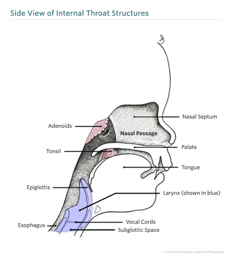
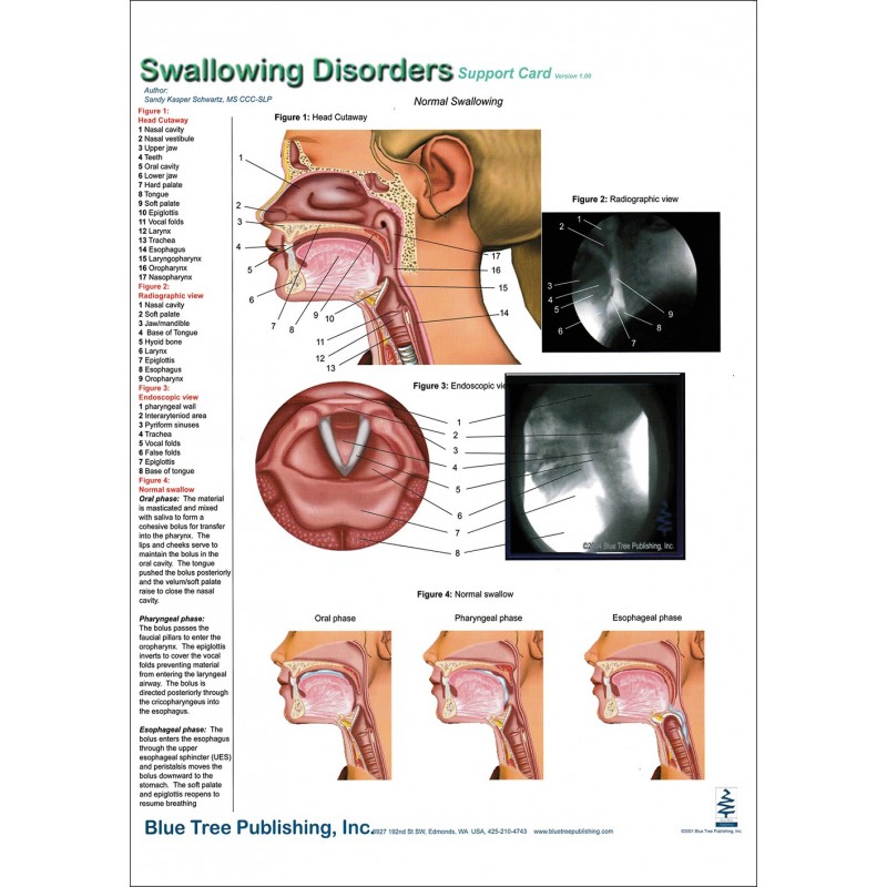
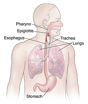
Anatomy of the Throat and Esophagus - Video & Lesson ... This is the muscular tube that connects the throat to the stomach. Your esophagus is about 10 inches long, and it acts only as a passageway for food and drink. The esophagus passes behind the ...
Throat diagram front view
The Front View of Male Reproductive System and More | New ... Male Reproductive System Front View. The male reproductive system consists of many different parts and structures which are all equally important. The task of the reproductive system in men is to produce male sex hormones and to make, preserve and carry the sperm as well as the semen. It is responsible for discharging sperm into the female ... The Eye's Drainage System, the Trabecular Meshwork ... The "angle" referred to here is the angle between the iris, which makes up the colored part of your eye, and the cornea, which is the clear-window front part of your eye. When the angle is open, your ophthalmologist can see most, if not all, of your eye's drainage system by using a special mirrored lens. Larynx anatomy: Cartilages, ligaments and muscles - Kenhub Larynx (anterior view) The larynx is a complex hollow structure located in the anterior midline region of the neck.It is anterior to the esophagus and at the level of the third to the sixth cervical vertebrae in its normal position. It consists of a cartilaginous skeleton connected by membranes, ligaments and associated muscles that suspend it from surrounding structures.
Throat diagram front view. Anatomy of a Dog's Throat - Cuteness.com A dog's throat anatomy is surprisingly similar to your own, starting with a dog esophagus and trachea and ending in the pup's stomach. Your dog may experience throat issues, including a collapsed trachea, so it's important to let your vet know if you've noticed anything unusual about his behavior. Human Body Diagrams Organs - Studying Diagrams The following human organ diagram shows you the front and back view of the human. Click on the labels below to find out more about your organs. ... In the Human Organs Library you can find pictures of eye ear mouth nose arm leg foot throat tooth stomach kidney liver lung heart intestine and vector bone structure of the. Anatomy, Thorax, Esophagus - StatPearls - NCBI Bookshelf The esophagus, historically also spelled oesophagus, is a tubular, elongated organ of the digestive system which connects the pharynx to the stomach. The esophagus is the organ that food travels through to reach the stomach for further digestion. It follows a path that travels behind the trachea and heart, in front of the spinal column, and through the diaphragm before entering the stomach.[1][2] Head and neck anatomy: Structures, arteries and nerves ... Head and neck (anterior view) The head and neck are two examples of the perfect anatomical marriage between form and function, mixed with a dash of complexity. The neck is resilient enough to sustain a five kilogram weight 24/7, yet sufficiently mobile to move it in several directions.
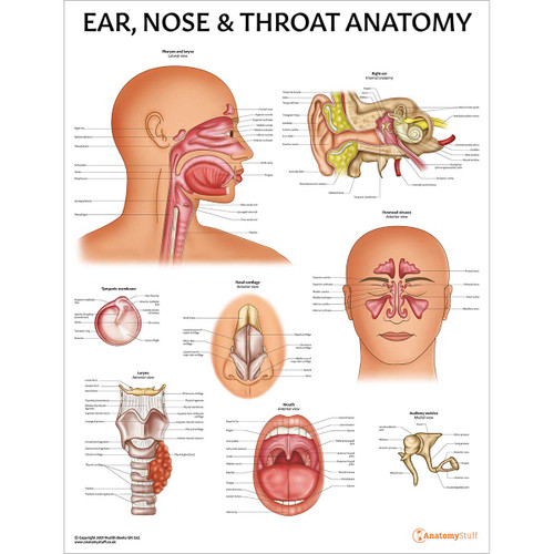
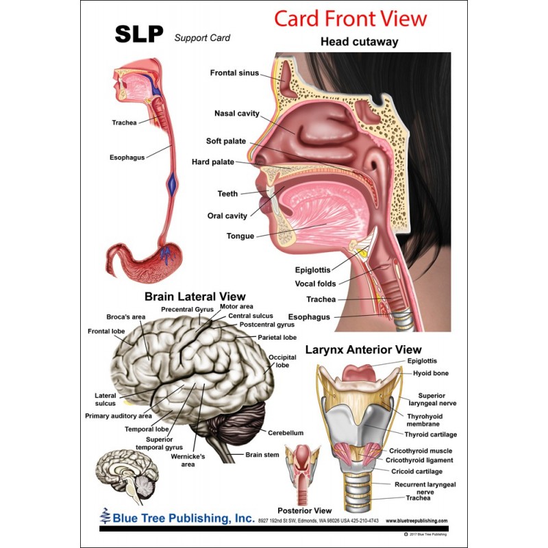
Anatomical landmarks adult - front view: MedlinePlus ... Anatomical landmarks adult - front view. Overview. There are three body views (front, back, and side) that can help you to identify a specific body area. The labels show areas of the body which are identified either by anatomical or by common names. For example, the back of the knee is called the "popliteal fossa," while the "flank" is ... Anatomy of the Throat and Neck | Dr. Larian The throat and neck anatomy consists of various structures, including: 1. Jugular Veins. There are four primary jugular veins: two internal and two external. Jugular veins drain blood from the neck, face, and brain and return it to the heart. The internal jugular veins are deep in the neck and the external jugular veins are immediately under ... How to Deep Throat: Everything You Need to Know ... - InStyle Also known as the pharyngeal reflex or laryngeal spasm, the gag reflex is the contraction of the back of the throat that occurs when triggered by an object touching the roof of the mouth, back of ... Throat Anatomy : Throat Parts, Pictures, Functions | HealthMD Parts Of A Throat. Based on anatomy, throat can be divided into 3 parts namely, the upper part, the middle part and the lower part called as nasopharynx, oropharynx and laryngopharynx respectively.. Pharynx - The elongated muscular tube connecting the back of the nose into the neck is known as pharynx.The function of pharynx is to transport the air from the nose into the larynx and to ...
Abdomen and digestive system diagrams: normal anatomy | e ... Full labeled anatomical diagrams - Anatomy of the abdomen and digestive system: these general diagrams show the digestive system, with the major human anatomical structures labeled (mouth, tongue, oral cavity, teeth, buccal glands, throat, pharynx, oesophagus, stomach, small intestine, large intestine, liver, gall bladder and pancreas). Neck Anatomy: Muscles, glands, organs - Kenhub Neck spaces. The content of the neck is grouped into 4 neck spaces, called the compartments.. Vertebral compartment: contains cervical vertebrae and postural muscles.; Visceral compartment: contains glands (thyroid, parathyroid, and thymus), the larynx, pharynx and trachea.; Two vascular compartments: contain the common carotid artery, internal jugular vein and the vagus nerve, on each side of ... Thorax: Anatomy, wall, cavity, organs & neurovasculature ... Thoracic wall The first step in understanding thorax anatomy is to find out its boundaries. The thoracic, or chest wall, consists of a skeletal framework, fascia, muscles, and neurovasculature - all connected together to form a strong and protective yet flexible cage.. The thorax has two major openings: the superior thoracic aperture found superiorly and the inferior thoracic aperture ... How to Do a Thyroid Neck Check - Verywell Health Stand in front of a mirror . Stand in front of a mirror so that you can see your neck. Be sure to remove any items, such as a scarf, necktie, jewelry, or turtleneck, that could block your view of your neck. If you use a hand-held mirror, direct it to focus on the lower-front portion of your neck.
Penis: 20 Different Types, Shapes, and Things to Know Positions that allow you to work the curve toward the front wall of the vagina or rectum give you the same hot-spot advantage as those with banana shapes. Pro tip: Try the T-bone.
Throat anatomy: MedlinePlus Medical Encyclopedia Image A.D.A.M., Inc. is accredited by URAC, for Health Content Provider ( ).URAC's accreditation program is an independent audit to verify that A.D.A.M. follows rigorous standards of quality and accountability. A.D.A.M. is among the first to achieve this important distinction for online health information and services.
Larynx anatomy: Cartilages, ligaments and muscles - Kenhub Larynx (anterior view) The larynx is a complex hollow structure located in the anterior midline region of the neck.It is anterior to the esophagus and at the level of the third to the sixth cervical vertebrae in its normal position. It consists of a cartilaginous skeleton connected by membranes, ligaments and associated muscles that suspend it from surrounding structures.
The Eye's Drainage System, the Trabecular Meshwork ... The "angle" referred to here is the angle between the iris, which makes up the colored part of your eye, and the cornea, which is the clear-window front part of your eye. When the angle is open, your ophthalmologist can see most, if not all, of your eye's drainage system by using a special mirrored lens.
The Front View of Male Reproductive System and More | New ... Male Reproductive System Front View. The male reproductive system consists of many different parts and structures which are all equally important. The task of the reproductive system in men is to produce male sex hormones and to make, preserve and carry the sperm as well as the semen. It is responsible for discharging sperm into the female ...






:background_color(FFFFFF):format(jpeg)/images/library/10807/Neck.png)











0 Response to "42 throat diagram front view"
Post a Comment