39 drag the labels onto the diagram to identify structural features associated with skeletal muscle.
Answered: Intercalated discs and pacemaker cells… | bartleby Q: Part B Drag the labels onto the diagram to identify structural features associated with the extrinsi... A: Introduction: a. Fibularis longus extends from proximal side of fibula and reaches first metatarsal... Identify the structures of skeletal muscle. - Brainly.com Feb 28, 2019 · Skeletal muscle is one of the major muscle types in most animals specialized to perform movement of the human body, maintaining body posture and generation of body heat present in different sizes and shapes. The main structure of skeletal muscle cells consists of various integrated tissues: 1.
similarities between christianity and judaism venn diagram Use pdf export for high quality prints and svg export for large sharp images or embed your diagrams anywhere with the creately viewer. Why educators should appear on-screen for instructional videos Drag the labels onto the diagram to identify structural features associated with skeletal muscle.
Drag the labels onto the diagram to identify structural features associated with skeletal muscle.
drag the appropriate labels to their respective targets ... Answer to drag the labels onto the diagram to identify structural features associated with skeletal muscle. A Coupled GCM-Cloud Resolving Modeling System, and A Regional Scale Model to Study Precipitation Processes. Insulin and glucagon act together to maintain homeostasis of blood glucose levels. Drag The Labels To Identify The Structures Of A Long Bone ... Sep 13, 2021 · Drag The Labels To Identify The Structures Of A Long Bone. / Drag The Labels Onto The Diagram To Identify Structural Features Associated With Skeletal Muscle Wiring Site Resource / Synchondroses are temporary joints which are only present in children, up until the end of puberty.. The skeletal system forms the framework of the body. Mastering A and P Assignment Unit 2 - Nervous System - StuDocu Drag the labels onto the diagram of neurochemical communication at an autonomic synapse. ANSWER: Correct. Art-labeling Activity Figure 11. Label the parts of the neuromuscular junction. Part A. Drag the labels onto the diagram to identify parts of the neuromuscular junction. ANSWER: Reset Help. Action potential arrives at varicosity.
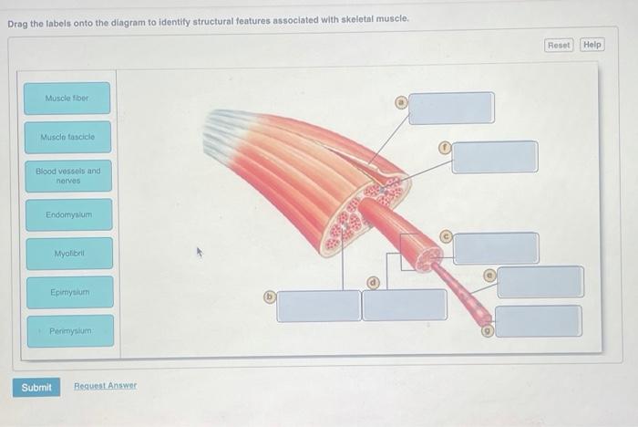
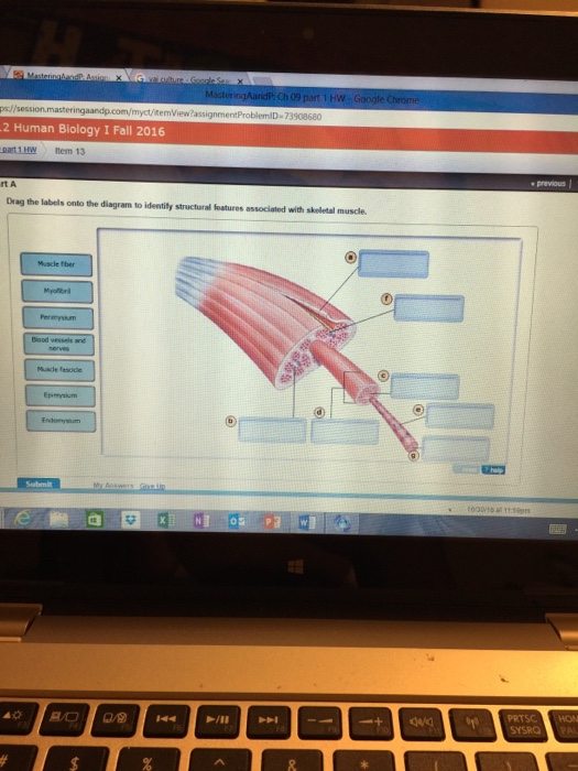
Drag the labels onto the diagram to identify structural features associated with skeletal muscle.. Ch 10 lab map Flashcards - Quizlet Drag the labels onto the diagram to identify structural features associated with a skeletal muscle fiber. look at pic Action potential propagation in a skeletal muscle fiber ceases when acetylcholine is removed from the synaptic cleft. Solved Drag the labels onto the diagram to identify | Chegg.com Features of Skeleton Muscle: The skeleton muscles are type of muscular tissue which attached with bones capable of voluntary… View the full answer Transcribed image text : Drag the labels onto the diagram to identify structural features associated with skeletal muscle. Mapping the Body | Boundless Anatomy and Physiology The Anterioposterior axis (AP axis) is the axis formed by the connection of the anterior (top) and posterior (bottom) ends of a region. The AP axis of a region is by definition perpendicular to the DV axis and vice-versa. The Left-to-right axis is the axis connecting the left and right hand sides of a region. Drag The Labels Onto The Diagram To Identify Structural ... Each skeletal muscle has three layers of connective tissue called mysia that enclose it and provide structure to the muscle as a whole and also compartmentalize the muscle fibers within the muscle. Drag the labels onto the diagram to identify structural features associated with skeletal muscle. The structure indicated by label e is part of ...
Drag The Labels Onto The Diagram To Identify Structural ... Drag the Labels Onto the Diagram to Identify Structural Features associated with Skeletal Muscle. solved drag the labels to the diagram to identify struc answer to drag the labels onto the diagram to identify structural features associated with skeletal muscle lecture exam 3 questions and study guide drag the labels onto the diagram to identify the processes and the structural ponents involved ... Skeletal Muscle Organization: Connective Tissue and Layers ... Skeletal muscle contains bundles of muscle fibers packed between layers of connective tissue called perimysium. Explore the role connective tissue plays in skeletal muscle organization and in ... (Get Answer) - Art-Labeling Activity: The Structure Of A ... Art-Labeling Activity: The Structure Of A Sarcomere Part A Drag The Labels To The Appropriate Location In The Figure. Reset Help A Band Barmere Hand Band MI Art-Labeling Activity: The Structure Of A Skeletal Muscle Fiber Part A Drag The Labels Onto The Diagram To Identity Structural Features Associated With A Skeletal Muscle Fiber. Synovial Joints | Anatomy and Physiology I - Lumen Learning The six types of synovial joints allow the body to move in a variety of ways. (a) Pivot joints allow for rotation around an axis, such as between the first and second cervical vertebrae, which allows for side-to-side rotation of the head. (b) The hinge joint of the elbow works like a door hinge. (c) The articulation between the trapezium carpal ...
lab 6 (exercises 12 and 13) Flashcards | Quizlet Drag the labels onto the diagram to identify structural features associated with skeletal muscle. Click card to see definition 👆. Tap card to see definition 👆. 1. epimysium. 2. perimysium. 3. endomysium. 4. nerve. 5. muscle fascicle. 6. muscle fibers. Drag The Correct Label To The Appropriate Location To ... Sarcoplasmic reiticulum and t tubules in the skeletal muscle fiber part a drag the appropriate labels to their respective targets. Terms in this set 6 haversian canal. Label the internal structure of a bone. Part a drag the correct label to the appropriate location to identify the features of a myofibril. 11.4 Identify the skeletal muscles and give their origins ... Editor's note: Replace figure with one that includes all muscles from table for example figure 10.7 from Marieb or 9.8 from Amerman. The orbicularis oris is a circular muscle that moves the lips, and the orbicularis oculi is a circular muscle that closes the eye. The occipitofrontalis muscle elevates the scalp and eyebrows. The muscle has a frontal belly and an occipital belly (near the ... chapter 10.pdf - chapter 10 chapter 10 Due 9:00am on ... Part A Drag the labels onto the diagram to identify structural features associated with skeletal muscle. contraction. secretion. conduction. cushioning. peristalsis. maintain posture maintain body temperature guard body entrances and exits produce movement All of the answers are correct.
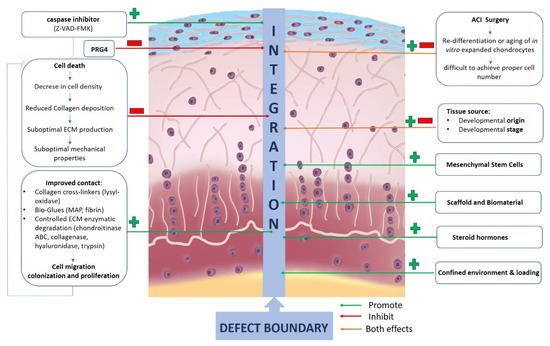
JFMK | Free Full-Text | Ex Vivo Systems to Study Chondrogenic ...
Answered: Label the muscles that move the… | bartleby A: In skeletal muscle contraction, the thin filaments are pulled which then slide past the thick filame... question_answer Q: Part B Drag the labels onto the diagram to identify structural features associated with the extrinsi...

Skeletal MyBP-C isoforms tune the molecular contractility of ...
Drag The Labels Onto The Diagram To Identify Structural ... 20 1 structure and function of blood vessels anatomy and physiology drag the labels onto the diagram to identify structural features associated with skeletal muscle. Each skeletal muscle has three layers of connective tissue called mysia that enclose it and provide structure to the muscle as a whole and also compartmentalize the muscle fibers ...
Associate Degree Nursing Physiology Review
Fibers And Hair Review Answers Worksheet [05N9IV] Part a drag the labels onto the diagram to identify structural features associated with skeletal muscle. Rigor mortis and algor mortis worksheets to practice determining PMI, due Thursday HW: guided notes for manner, cause & mechanism of death, types of injuries, and pre/post mortem injuries. Causes of the american revolution worksheet answers.
Neural Control of Human Movement
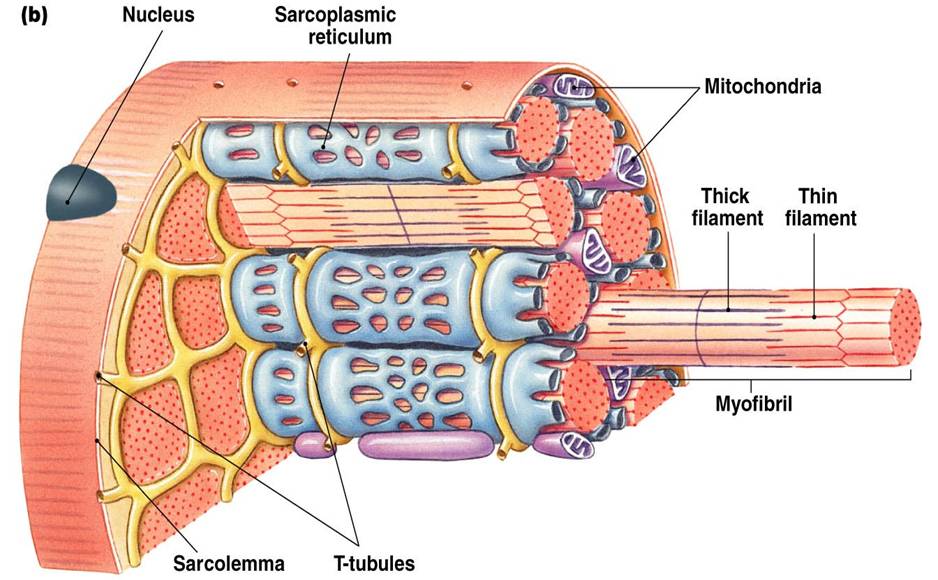
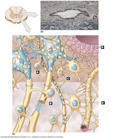
PDF CHAPTER 4: TISSUES - Warner Pacific University Location: In skeletal muscles attached to bones or occasionally to skin. Photomicrograph: Skeletal muscle (approx. 460x). Notice the obvious banding pattern and the fact that these large cells are multinucleate. Nuclei Striations Part of muscle fiber (cell)

Enhanced single-cell encapsulation in microfluidic devices ...
Part a muscle tissue has the ability to ... - Course Hero Correct Art-labeling Activity: The Organization of Skeletal Muscles (1 of 2) Drag the labels onto the diagram to identify structural features associated with skeletal muscle. Part A Drag the labels onto the diagram to identify structural features associated with skeletal muscle.
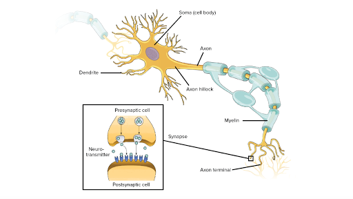
Overview of neuron structure and function (article) | Khan ...
Drag The Labels Onto The Diagram To Identify Structural ... Drag the labels onto the diagram to identify structural features associated with a skeletal muscle fiber. look at pic action potential propagation in a skeletal muscle fiber ceases when acetylcholine is removed from the synaptic cleft.

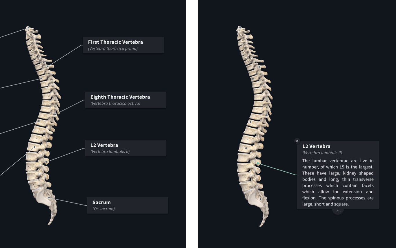
The Skeletal System: Labelling the Bones - ppt video online ...
10.2 Skeletal Muscle - Anatomy & Physiology Each skeletal muscle has three layers of connective tissue that enclose it, provide structure to the muscle, and compartmentalize the muscle fibers within the muscle (Figure 10.2.1). Each muscle is wrapped in a sheath of dense, irregular connective tissue called the epimysium , which allows a muscle to contract and move powerfully while ...
Answered: Drag the labels onto the diagram to… | bartleby
Schon Respiratory System With Labels Drag the labels onto the diagram to identify structural features associated with skeletal muscle. This respiratory tree ends in puffy structures called alveoli that are made of a single layer of squamous cells surrounded by a network of capillaries. SEE ALSO : Frisch N64 Top Labels

Practices for Measuring 3D Organelle Morphology and ...
Drag the labels on the left onto the diagram Part a drag the labels onto the diagram to identify structural features associated with skeletal muscle. This is the recommended approach, because manual layout allows you to precisely position elements where you want them, consistently across multiple diagrams.

art A rag the labels onto the diagram to identify the parts ...
28 Eaton Lighting Contactor Wiring Diagram - Diagram ... 30a power pole rating up to 12 poles maximum power poles latch easily onto the base and. Collection of eaton lighting contactor wiring diagram. Eaton Motor Starter Wiring Diagram - impremedia.net A wiring diagram is a streamlined standard photographic depiction of an electrical circuit.
Solved Drag the labels onto the diagram to identity | Chegg.com
Mastering A and P Assignment Unit 2 - Nervous System - StuDocu Drag the labels onto the diagram of neurochemical communication at an autonomic synapse. ANSWER: Correct. Art-labeling Activity Figure 11. Label the parts of the neuromuscular junction. Part A. Drag the labels onto the diagram to identify parts of the neuromuscular junction. ANSWER: Reset Help. Action potential arrives at varicosity.

10.2 Skeletal Muscle – Anatomy & Physiology
Drag The Labels To Identify The Structures Of A Long Bone ... Sep 13, 2021 · Drag The Labels To Identify The Structures Of A Long Bone. / Drag The Labels Onto The Diagram To Identify Structural Features Associated With Skeletal Muscle Wiring Site Resource / Synchondroses are temporary joints which are only present in children, up until the end of puberty.. The skeletal system forms the framework of the body.
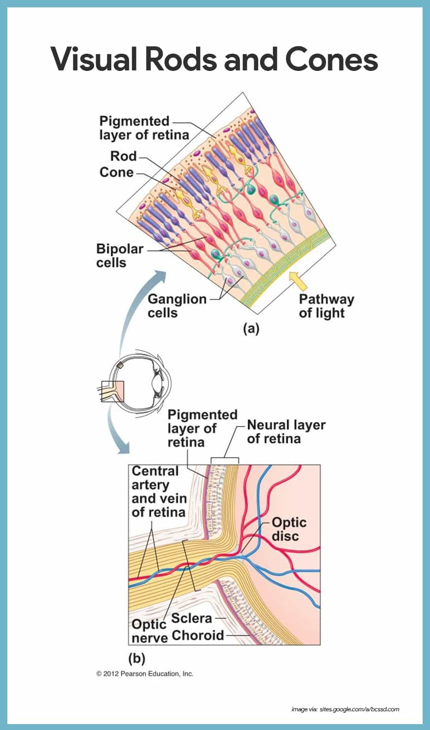
Special Senses Anatomy and Physiology - Nurseslabs
drag the appropriate labels to their respective targets ... Answer to drag the labels onto the diagram to identify structural features associated with skeletal muscle. A Coupled GCM-Cloud Resolving Modeling System, and A Regional Scale Model to Study Precipitation Processes. Insulin and glucagon act together to maintain homeostasis of blood glucose levels.
Solved Drag the labels onto the diagram to identify | Chegg.com
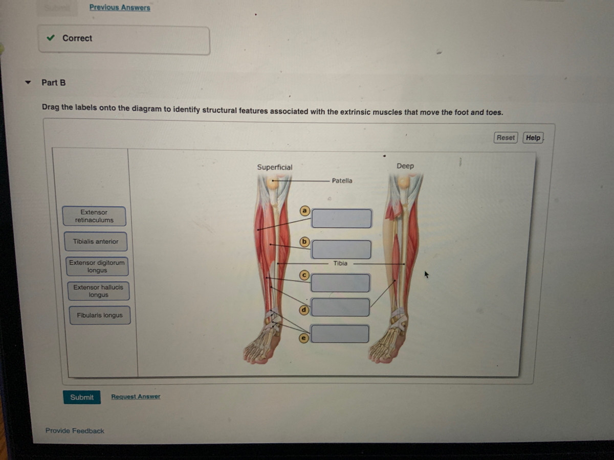
Answered: Part B Drag the labels onto the diagram… | bartleby

Teruo Okano's 193 research works in Biology and Agricultural ...

Drag the labels onto the diagram to identify parts of the ...

Art Labeling Activity Levels Of Protein Structure at Level
Overview of the Muscular System | Boundless Anatomy and ...

FINAL A&P Chapter 9 & 11 Flashcards | Quizlet

Chapter 9: Muscles and Muscles Tissue | Muscle diagram ...
Kinesiology
Chapter 12-Neural Tissue Flashcards - Easy Notecards
MUSCLES

Actin Residue Glu93 Is Identified as an Amino Acid Affecting ...
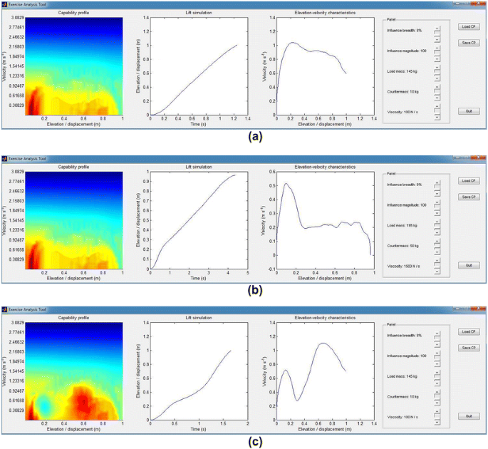
Computer-Aided Parameter Selection for Resistance Exercise ...
Labels | Complete Anatomy
Pancreas & Insulin
POLITECNICO DI TORINO

Pfam: Family: Fer2_3 (PF13085)
Frontiers | Influence of the Mechanical Environment on the ...

Ch 10 lab map Flashcards | Quizlet

Skeletal MyBP-C isoforms tune the molecular contractility of ...

Exercise 6 Review Sheet Art-labeling Activity 5 Drag the ...

1.4 The Somatic Nervous System – Neuroscience: Canadian 1st ...
Muscles and Muscle Tissue

UCSC Genome Browser: News Archives

lab 6 (exercises 12 and 13) Flashcards | Quizlet

Part A Muscle tissue has the ability to contract when ...
0 Response to "39 drag the labels onto the diagram to identify structural features associated with skeletal muscle."
Post a Comment