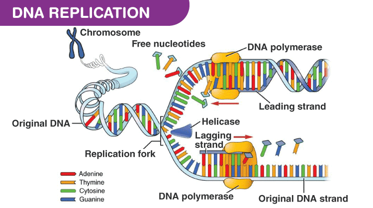
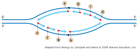
42 the diagram below shows a bacterial replication fork and its principal proteins.
Saccharomyces cerevisiae - an overview | ScienceDirect Topics Saccharomyces cerevisiae is the principal yeast utilized in biotechnology worldwide, due largely to its unique physiology and associated key roles in many food fermentations and other industrial processes (Phaff et al. DNA Replication Steps and Process DNA replication is the process of copying the DNA within our cells. This process involves RNA Once completed, the parent strand and its complementary DNA strand coils into the familiar double helix shape. It forms the replication fork by breaking hydrogen bonds between nucleotide pairs in DNA.
The Diagram Below Shows A Bacterial Replication Fork And Its... The diagram below shows a bacterial replication fork and its principal proteins. The parental dna is shown in dark blue the newly synthesiz...
The diagram below shows a bacterial replication fork and its principal proteins.
The Molecular Basis of Inheritance | PDF | Dna Replication | Primer... DNA replication always begins at an origin of replication. In bacteria, there is a single origin of replication on the Drag the arrows onto the diagram below to indicate the direction that DNA polymerase III moves along the This image shows a replication bubble in a bacterial chromosome. The diagram below shows a bacterial replication fork and its... Drag the labels to their appropriate locations in the diagram to describe the name or function of each structure. Use pink labels for the pink targets and blue labels for the blue targets. Answer. e. lagging f. leading (a) breakes hydrogen bonds, unwinding DNA double helix. Molecular Expressions Cell Biology: Bacteria Cell Structure Bacteria give yogurt its tangy flavor and sourdough bread its sour taste. They make it possible for ruminant animals (cows, sheep, goats) to digest Bacteria are prokaryotes, lacking well-defined nuclei and membrane-bound organelles, and with chromosomes composed of a single closed DNA circle.
The diagram below shows a bacterial replication fork and its principal proteins.. Bacterial Replication Fork Diagram Answers - Free Catalogs A to Z The Diagram Below Shows A Bacterial Replication Fork And. 7 hours ago The diagram below shows a replication fork with the two parental DNA strands labeled at their 3' and 5 4/4 (5). The diagram below shows a bacterial replication fork and its principal proteins. DNA replication - Wikipedia A number of proteins are associated with the replication fork to help in the initiation and continuation of DNA synthesis. For a cell to divide, it must first replicate its DNA.[18] DNA replication is an all-or-none process; once replication begins, it proceeds to completion. The Diagram Below Shows A Bacterial Replication Fork And Its... ...replication fork and its principal proteins, Drag the labels to their appropriate locations in the Traction the brand to their ideal locations in the diagram to explain the surname or duty of every Number of proteins are affiliated in DNA replication, consisting of the following: Helicase division the... 31 The Diagram Below Shows A Bacterial Replication Fork And Its... The diagram below shows a replication bubble with synthesis of the leading and lagging strands on both sides of the bubble. Replication Fork Breakage And Restart In Escherichia Coli. The Diagram Below Shows A Bacterial Crispr Cas Dna Supercoiling And Nucleoid Associated Proteins.
The Diagram Below Shows A Bacterial Replication Fork And Its... The diagram below shows a replication fork with the two parental DNA strands labeled at their 3' and 5 4/4(5). The diagram below In this tutorial you will learn how DNA is replicated and understand the roles of the proteins involved in the process. Part A The mechanism of DNA replication The... DNA replication is carried out by a complex system of enzymes Single Stranded Binding (SSB) Proteins. SSB proteins bind to the DNA strands after they have been separated and prevent the strands from re-annealing. On the leading strand, DNA pol III is moving towards the replication fork and can synthesise continuously. Different Size, Shape and Arrangement of Bacterial Cells Different Size, Shape and Arrangement of Bacterial Cells. When viewed under light microscope, most bacteria appear in variations of three major shapes: the rod (bacillus), the sphere (coccus) and the spiral type (vibrio). Changing the way you learn | Quiz d. Its use is incompatible with the goals of domain modeling. e. The diagrams require you to include too much detail. a) A component diagram shows a configuration of the system at a given moment. b) A component diagram shows all potential interactions among components in a view.
The Diagram Below Shows A Bacterial Replication Fork And Its... Mastering biology exam 3. The diagram below shows a bacterial replication fork and its principal proteins. Solved I Am New... What are bacteria? | Live Science Bacteria are microscopic single-celled organisms that can be helpful, such as those that live in our guts, or harmful, such as flesh-eating bacteria. Live Science is supported by its audience. When you purchase through links on our site, we may earn an affiliate commission. DNA Replication | Microbiology DNA replication uses a large number of proteins and enzymes (Table 1). Table 1. The Molecular Machinery Involved in Bacterial DNA Replication. Figure 3. This structure shows the guanosine triphosphate deoxyribonucleotide that is incorporated into a growing DNA strand by cleaving the two... Details: The diagram below shows a bacterial replication fork and... Bacterial Replication Fork Drivers! find and download drivers laptops, computer, printer for windows, mac. Once the strands are separated, a group of proteins called helper … bacterial replication video. Details: Processes occurring at a bacterial replication fork DNA replication requires three...
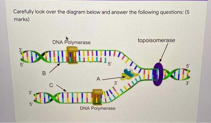
Solved Carefully look over the diagram below and answer the ...
Bacillus subtilis - microbewiki | Chromosome Replication Replication proceeds bidirectionally and two replication forks progress in the clockwise and counterclockwise directions along the chromosome halves. They also showed a significant increase in duodenal and jejunal protease and trypsin activities and a decrease in pancreatic trypsin activity.
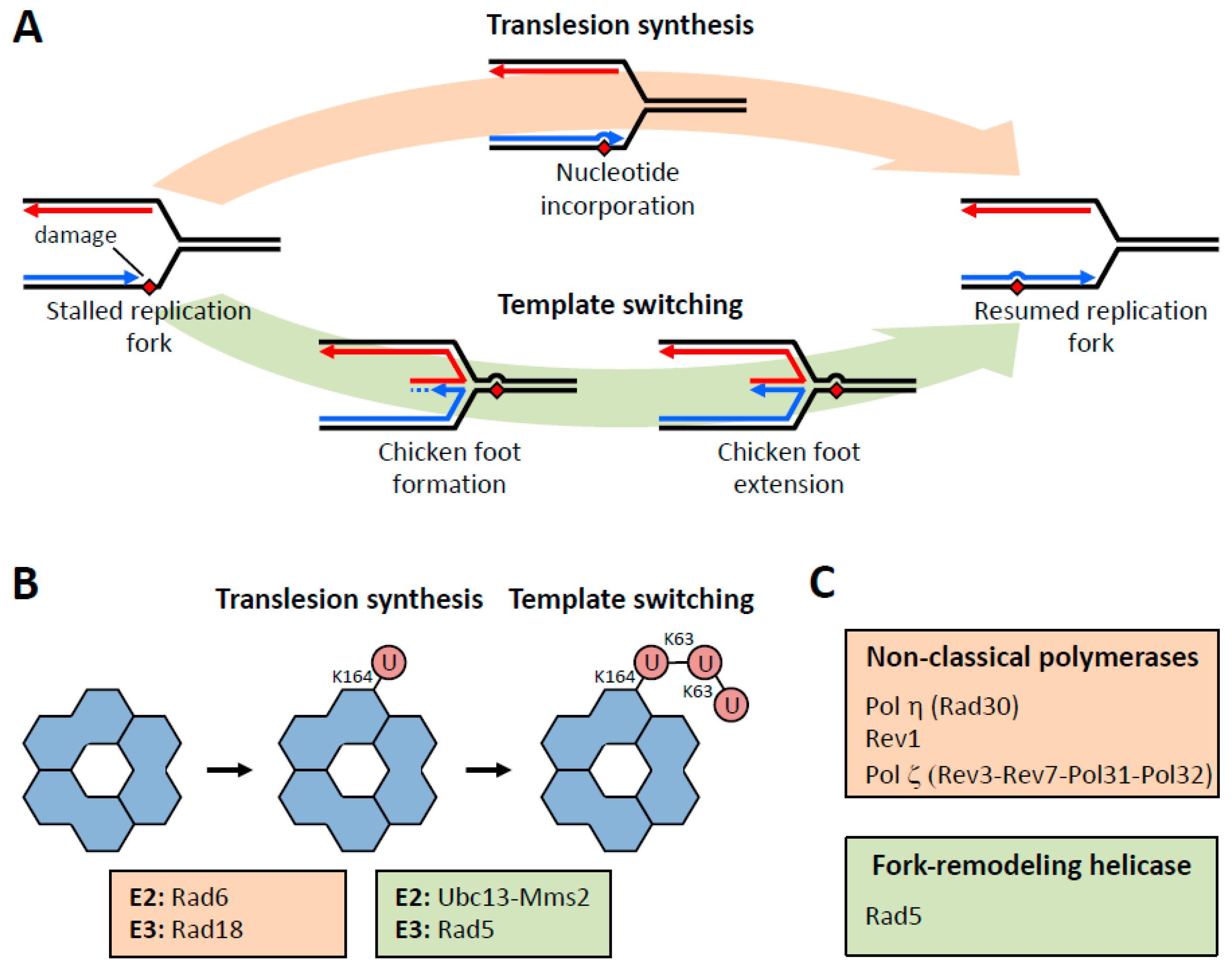
Genes | Free Full-Text | Control of DNA Damage Bypass by ...
BREAKING - Three studies published by the CDC, UK Government... She writes that since the principal reason of a mandate is to protect others from infection, and these studies prove beyond a shadow of a doubt that they do not do this, those who mandate the Covid-19 injections may wish to seek legal counsel regarding their culpability and liability for potential...
PCR and Molecular Biology Fundamental Principles
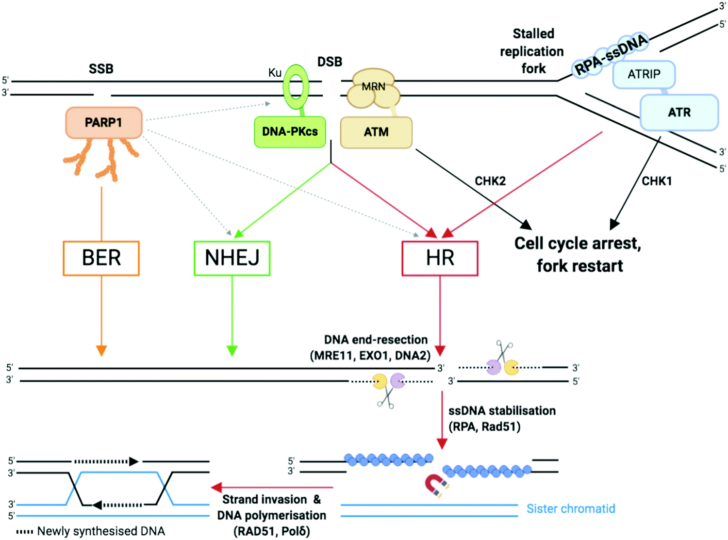
Nucleases and Co-Factors in DNA Replication Stress Responses (B) Diagram of a replication fork in which the leading strand DNA polymerase is blocked and decouples from the MCM helicase, creating ssDNA that is bound by RPA. ATRIP recognition of ssDNA-RPA recruits and activates ATR. (C) Crosstalk among phosphatidyl inositol 3′ kinase-related kinases...
Solved Part B Processes occurring at a bacterial replication ...
The Diagram Below Shows A Bacterial Replication Fork And Its... The diagram below shows a replication fork with the two parental dna strands labeled at their 3 and 5 ends. Use pink labels for the pink targets and blue labels Nucleoid Occlusion Protein Noc Recruits Dna To The Bacterial Cell. Guidelines For Dna Recombination And Repair Studies Cellular Assays.
Bio 102 Practice Problems Chromosomes and DNA Replication
New studies show that COVID vaccines damage your immune system... The results show what is really happening, and Nobody has been able to attack the paper with a credible argument, even on Twitter.

Replisome - an overview | ScienceDirect Topics
Genetics Flashcards | Quizlet Part B - Processes occurring at a bacterial replication fork The diagram below shows a bacterial replication fork and its principal proteins. Drag the labels to their appropriate locations in the diagram to describe the name or function of each structure.

Processing ribonucleotides incorporated during eukaryotic DNA ...
Bacterial Replication Termination - Proteopedia, life in 3D Replication is terminated in bacterial systems such as E.coli and B.subtilis by a "replication fork trap" Mutational analysis a contrahelicase region has shown that mutations within these regions abolish The role of the replication fork arrest was believed to be of great importance for the faithful...
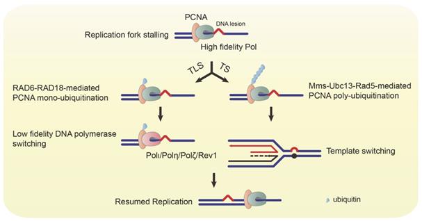
Post-Translational Modifications of PCNA in Control of DNA ...
Solved The diagram below shows a bacterial replication fork Transcribed image text : The diagram below shows a bacterial replication fork and its principal proteins. Drag the labels to their appropriate locations in the diagram to describe the name or function of each structure. Use pink labels for the pink targets and blue labels for the blue targets.

PCNA ubiquitination protects stalled replication forks from ...
How SDS-PAGE Works: 7 Key Points Every Scientist Should Know This is shown in the diagram below. Control of the charge state of the glycine by the different buffers is the key to the whole stacking gel thing. All of the proteins in the gel sample have an electrophoretic mobility that is intermediate between the extreme of the mobility of the glycine and Cl...

PDF) Division of Labor between PCNA Loaders in DNA ...
Proteins- Definition, Properties, Structure, Classification, Functions Proteins- Properties, Structure, Classification and Functions. Proteins are the polymers of amino acids covalently linked by the peptide bonds. If a denatured protein returns to its native state after the denaturing agent is removed, the process is called renaturation.

Chapter 16 BSC 2010 Flashcards | Quizlet
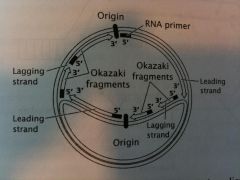
Exam 3: Chs. 5 (DNA Structure and Replication...) - Easy Notecards 5 (DNA Structure and Replication Machinery) & 16 (The Molecular Basis of Inheritance) Flashcards. Below is an example of a DNA sequence and its complement. This image shows a replication bubble in a bacterial chromosome. The region enclosed by the box includes the two Drag the arrows onto the diagram below to indicate the direction that DNA polymerase III moves along the parental...
DNA folds threaten genetic stability and can be leveraged for ...
IELTS Process Diagram - How To Write a Process Essay - IELTS... An IELTS process diagram question can contain a wide variety of different types of graphics. It could be a natural process such as the water cycle, a manufacturing process or a Here's our practice question: The diagrams below show a structure that is used to generate electricity from wave power.
DNA Replication Process with Diagrams Class 12 - Prokaryotic ...
Molecular Expressions Cell Biology: Bacteria Cell Structure Bacteria give yogurt its tangy flavor and sourdough bread its sour taste. They make it possible for ruminant animals (cows, sheep, goats) to digest Bacteria are prokaryotes, lacking well-defined nuclei and membrane-bound organelles, and with chromosomes composed of a single closed DNA circle.
Bio 102 Practice Problems Chromosomes and DNA Replication
The diagram below shows a bacterial replication fork and its... Drag the labels to their appropriate locations in the diagram to describe the name or function of each structure. Use pink labels for the pink targets and blue labels for the blue targets. Answer. e. lagging f. leading (a) breakes hydrogen bonds, unwinding DNA double helix.

PCR and Molecular Biology Fundamental Principles
The Molecular Basis of Inheritance | PDF | Dna Replication | Primer... DNA replication always begins at an origin of replication. In bacteria, there is a single origin of replication on the Drag the arrows onto the diagram below to indicate the direction that DNA polymerase III moves along the This image shows a replication bubble in a bacterial chromosome.
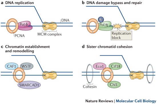
Regulation of PCNA–protein interactions for genome stability ...
Mastering Biology Chapter 16 – RHS Homework

PDF) A Link between Replicative Stress, Lamin Proteins, and ...

Mastering Biology Chp. 13 HW Flashcards | Quizlet
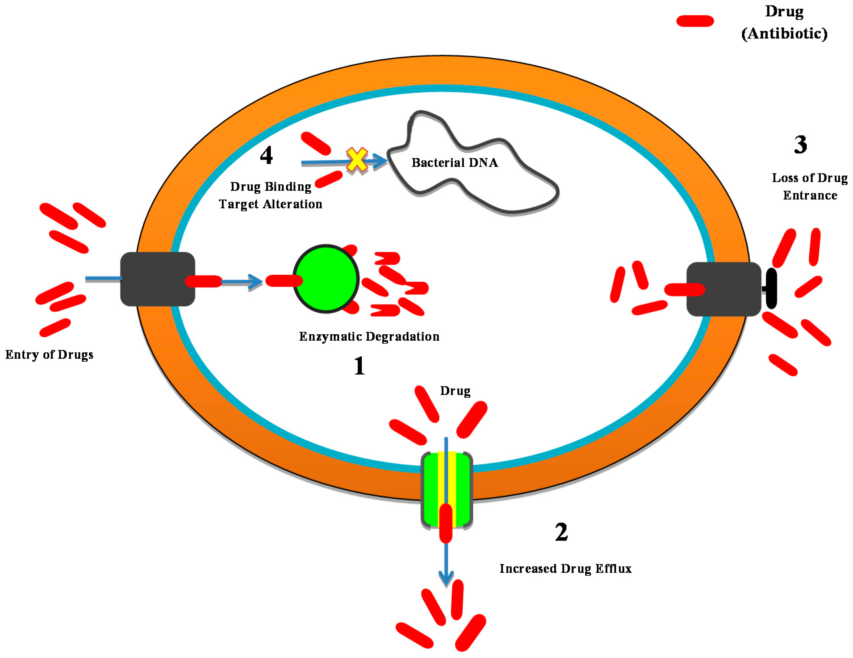
Antibiotics | Free Full-Text | Nanotechnology as a Novel ...
Mastering Biology Chapter 16 – RHS Homework
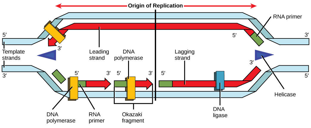
9.2 DNA Replication – Concepts of Biology – 1st Canadian Edition
Genes | Free Full-Text | Subcellular Dynamics of a Conserved ...

PDF) A Family Portrait of the RIO kinases

Replisome Dynamics and Their Functional Relevance upon DNA ...

DNA double-strand break repair-pathway choice in somatic ...

The E. coli DNA Replication Fork - ScienceDirect

94 CIE biology ideas | biology, teaching biology, biology ...

Mastering Biology ch. 13 Flashcards | Quizlet

Mastering Biology Chapter 16 – RHS Homework
DNA double-strand break repair-pathway choice in somatic ...
Frontiers | The DnaA Cycle in Escherichia coli: Activation ...

DNA replication of prokaryotes
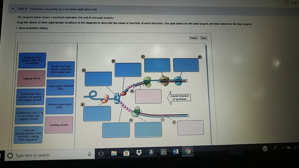
Solved Part B Processes occurring at a bacterial replication ...
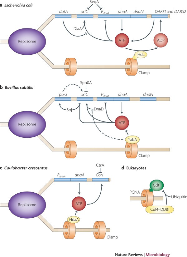
Regulation of the replication cycle: conserved and diverse ...
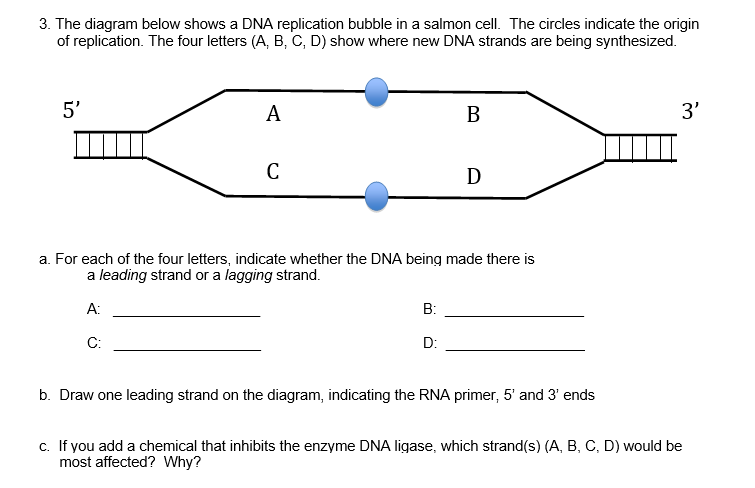
Solved The diagram below shows a DNA replication bubble in a ...

Mastering Biology Exam 3 Flashcards | Quizlet

Major Enzymes | Biology for Majors I
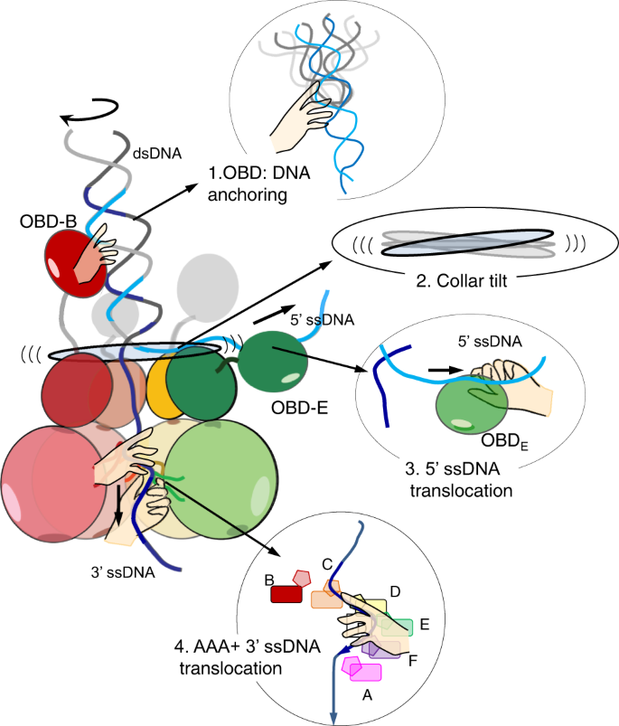
Unwinding of a DNA replication fork by a hexameric viral ...

Deciphering UV‐induced DNA Damage Responses to Prevent and ...

intro to cell test five Flashcards & Practice Test | Quizlet
Genetics Chap 12, 13, 14, & 15 Flashcards - Cram.com
0 Response to "42 the diagram below shows a bacterial replication fork and its principal proteins."
Post a Comment