37 brain and spinal cord diagram
Spinal Cord Brain Diagram. angelo. December 1, 2021. Brain And Spinal Cord Anatomy From The Merck Manual Home Healthbook Basic Anatomy And Physiology Brain Anatomy Medical Anatomy. Find Hd Draw A Labelled Diagram Of A Section Of Human Brain Human Brain Class 10 Hd Png Download To Search And Download Mor Human Brain Brain Diagram Brain. (11) This brain part controls involuntary actions such as breathing, heartbeats, and digestion. (12) cerebrum cerebellum brain stem spinal cord This part of the nervous system moves messages between the brain and the body.
In the brain, white matter is buried under the grey surface, carrying signals across different parts of the brain. In the spinal cord, white matter is the external layer surrounding the grey core. The brain. Image: QBI/Levent Efe. If the CNS is the processing centre of the human body, the brain is its headquarters.
Brain and spinal cord diagram
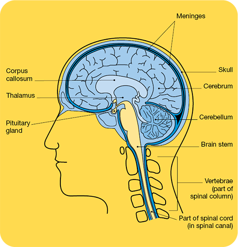
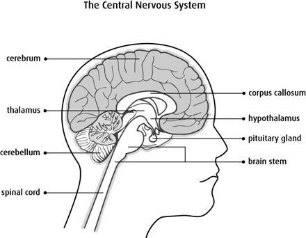
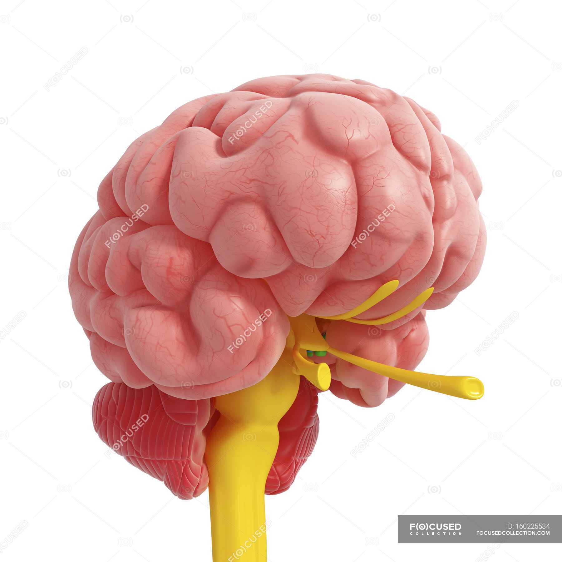
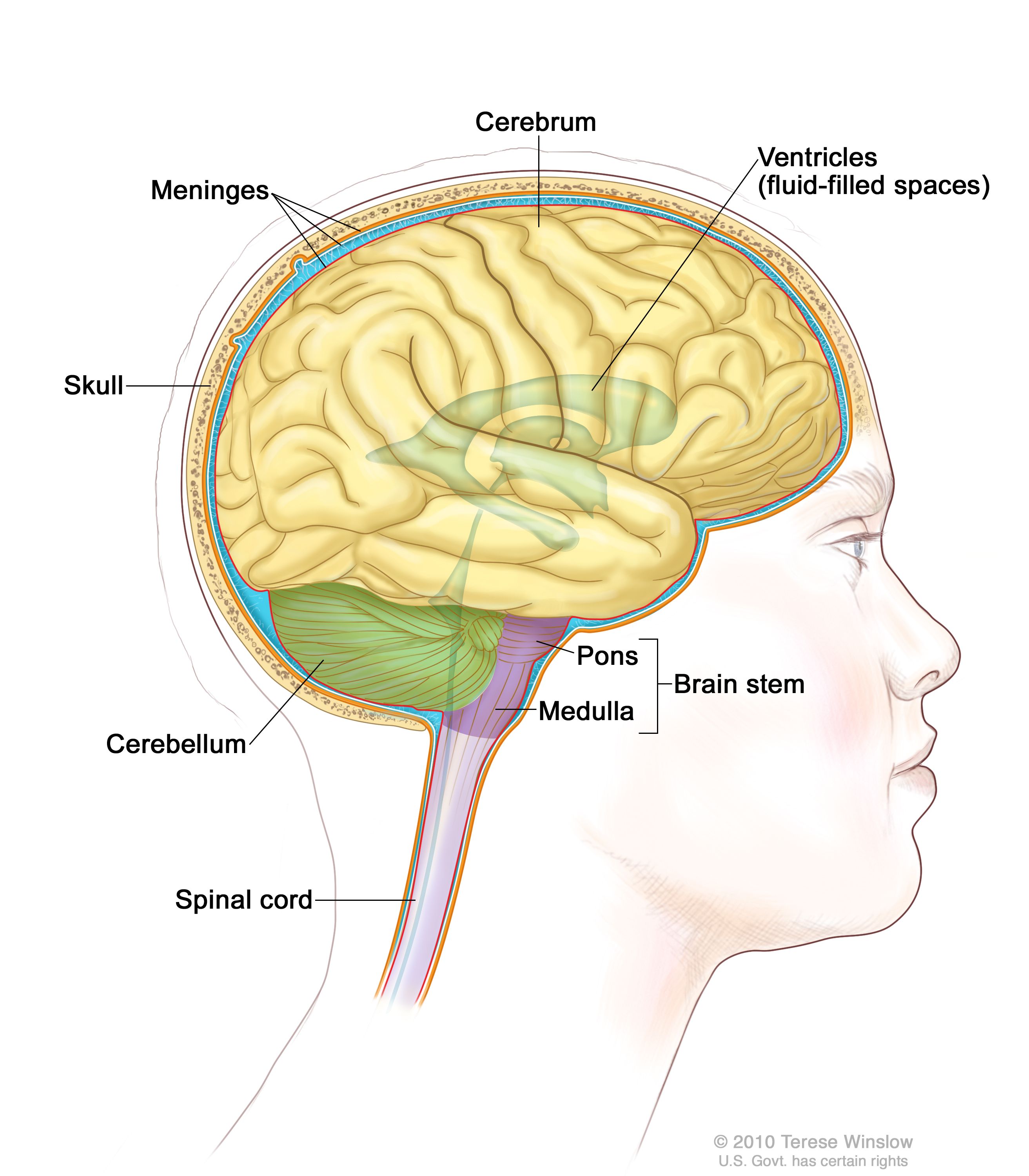
The spinal cord is a single structure, whereas the adult brain is described in terms of four major regions: the cerebrum, the diencephalon, the brain stem, and the cerebellum. A person's conscious experiences are based on neural activity in the brain. The regulation of homeostasis is governed by a specialized region in the brain. Spinal Cord Diagram. The spinal cord is one of the most important structures in the human body. In fact, it is the most important structure for any vertebrates. Anatomically, the spinal cord is made up is made up of nervous tissue and is integrated into the spinal column of the backbone. The spinal cord consists of ascending and descending tracts.The ascending tracts are sensory pathways that travel through the white matter of the spinal cord, carrying somatosensory information up to the brain.They allow you to feel sensations from the external environment (exteroceptive) such as pain, temperature, touch, as well as proprioceptive information from muscles and joints.
Brain and spinal cord diagram. Start studying The Nervous System: Brain and Spinal Cord. Learn vocabulary, terms, and more with flashcards, games, and other study tools. Ventricles are hollow cavities of the brain, that contain the cerebrospinal fluid (CSF), which circulates within the brain and spinal cord. There are all together four ventricles in the human brain, that constitute the ventricular system, along with the cerebral aqueduct. They are known as, lateral ventricles, third ventricle, and fourth ventricle. The spinal cord is active when the brain is busy. It is also called the 2 nd brain of the human body. It begins in continuation with the medulla oblongata and extends; The spinal cord is enclosed in a bony cage called vertebral; A total of thirty-one pairs of spinal nerves arise from the spinal; Functions of Spinal Cord • The brain stem is between the spinal cord and the rest of the brain. Basic functions like breathing and sleep are controlled here. • The basal ganglia are a cluster of structures in the ...
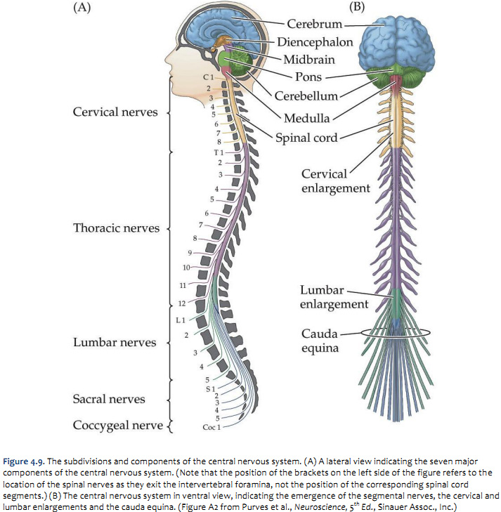
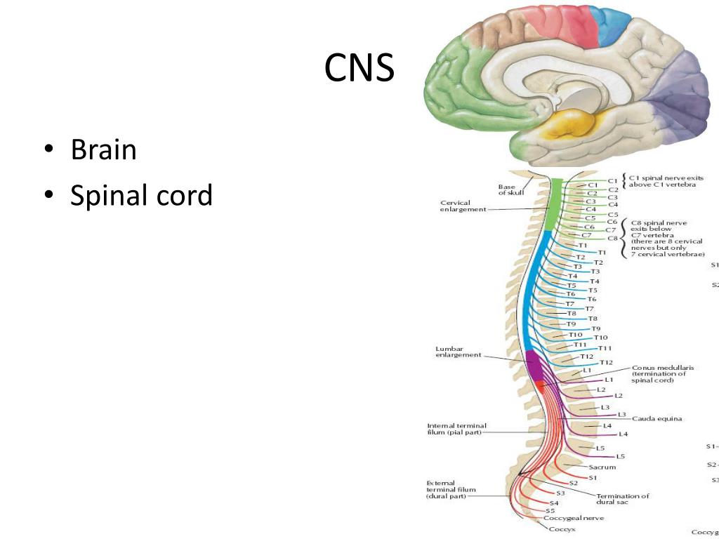
A complete spinal cord injury means that there is a total blockage of signals from the brain to your sacral nerves. An incomplete spinal cord injury means there is some preservation of nerves from the brain to the lowest part of the spinal cord, the sacral level. The amount of movement and feeling that This is an online quiz called Label Parts of the Brain. There is a printable worksheet available for download here so you can take the quiz with pen and paper. From the quiz author. Okay. ... Label the Brain and Spinal Cord 10p Image Quiz. PurposeGames Create. Play. Learn. The brain stem begins inferior to the thalamus and runs approximately 7 cm before merging into the spinal cord. The brain stem centers produce the rigidly programmed, automatic behaviors necessary for survival. Positioned between the cerebrum and the spinal cord, the brain stem also provides a pathway for fiber tracts ... Spinal nerves (diagram) Spinal nervesemerge from the segments of the spinal cord. They are numbered according to their specific segment of origin. Hence, the 31 pairs of spinal nerves are divided into 8 cervical pairs, 12 thoracic pairs, 5 lumbar pairs, 5 sacral pairs, and 1 coccygeal spinal nerve. All spinal nerves are mixed, containing both ...
lower-motor neuron in the brain stem or spinal cord. The axon of the lower-motor neuron has direct control over skeletal muscle fibers. Stimulation of the lower- motor neuron always has an excitatory effect on the skeletal muscle fibers. Skeletal muscle Skeletal muscle Somatic motor nuclei of brain stem Lower motor neurons Connecting the brain to the spinal cord, the brainstem is the most inferior portion of our brain. Many of the most basic survival functions of the brain are controlled by the brainstem. The brainstem is made of three regions: the medulla oblongata, the pons, and the midbrain. Human Spinal Cord: Structure and Effects (With Diagram) The human spinal cord is made up 31 segments. From each of these segments, a pair of spinal nerves takes origin. Hence there are 31 pairs of spinal nerves. Of 31 pairs: i. 8 belong to cervical segments. ii. 12 belong to thoracic segments. The spinal motor neurons project out of the cord to the correct muscles via the ventral root. These connections control conscious movements, such as writing and running. Information also flows in the opposite direction resulting in involuntary movement. Sensory neurons provide feedback to the brain via the dorsal root.
The spine diagram shown below, consists of many bones or vertebrae,soft discs,the spinal cord, and spinal nerves. The spine anatomy is a complex structure. Simply put: the vertebrae, which stack like spools of thread, support the back and protect the spinal cord. In turn, the spinal cord relays essential information between the brain and the body.

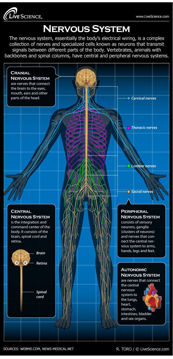
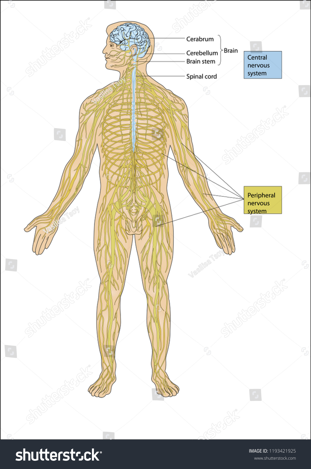
The extension of the central nervous system is through the medulla oblongata, which continues as the spinal cord, by exiting the skull through the foramen magnum and continuing in the spinal column. This is where the spinal cord gives out nerves which form the peripheral nervous system.

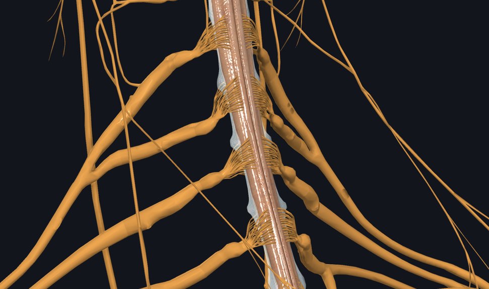
The spinal cord is part of the central nervous system and serves as a kind of superhighway. Sensory information and motor commands travel up and down, heading to and from the brain. These signals speed in and out of the spinal cord via spinal nerves—the "on-ramps and off-ramps" that branch out to supply the limbs, torso, and pelvis. Some ...
Brain And Spinal Cord Diagram Name angelo. November 28, 2021. Pin On Optometry . ... Nervous System For Kids Brain Spinal Cord Nerves Human Body Nervous System Human Nervous System Human Body Unit . In cord, diagram, name. Leave a Reply Cancel reply. You must be logged in to post a comment.
This video discuss the anatomy of the spine. It is part of the DVD series "Understanding Spinal Cord Injury" created by Shepherd Center. Visit spinalinjury10...
The brain stem connects the spinal cord to the higher-thinking centers of the brain. It consists of three structures: the medulla oblongata, the pons, and the midbrain. The medulla oblongata is continuous with the spinal cord and connects to the pons above. Both the medulla and the pons are considered part of the hindbrain.
Function: Connects Vertebro-basilar system and Internal carotid system. Circle of Willis (Function) 1) To maintain COLLATERAL CIRCULATION in case of blockage of any of the components. 2) Equalizes blood flow to different parts of the brain. 3) Connects Vertebro-basilar system and Internal carotid system. Circle of Willis Defects.
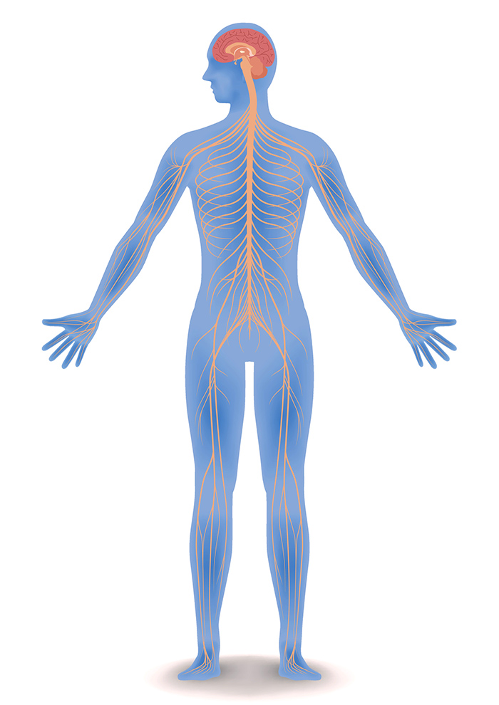
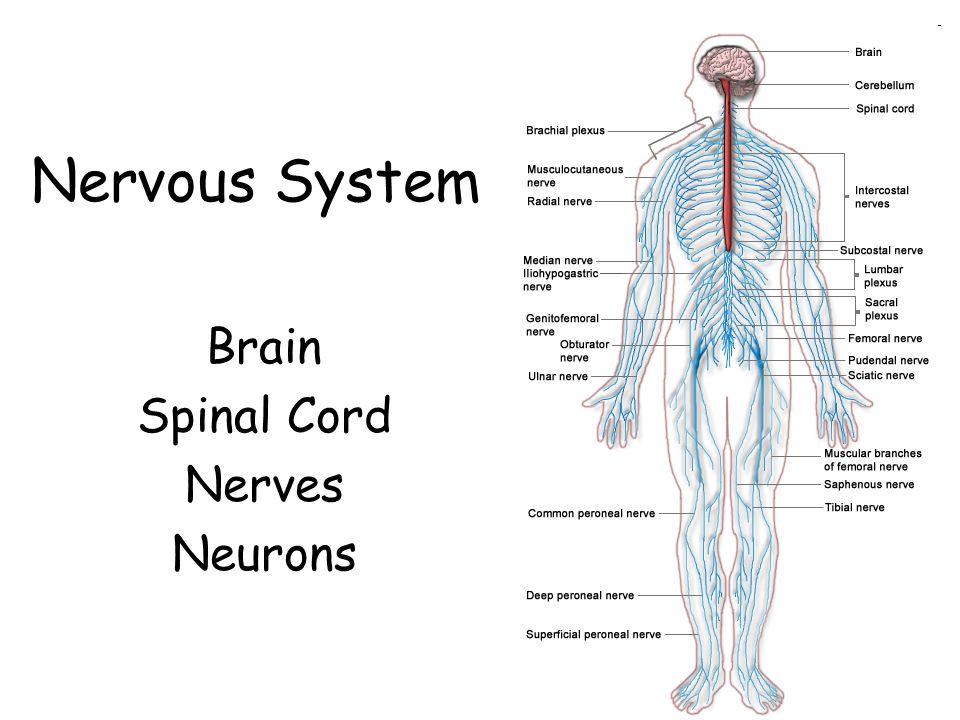
Together with the brain and the spinal cord, the nerves constitute the nervous system. To distinguish the control centres from the information pathways, we divide the nervous system into two sub-systems (to see them, run your cursor over their names in the following diagram):
Spinal Cord Controls simple reflexes Pathway to neural fibers Medulla Controls/regulates heartbeat and breathing To and from brain Reticular Formation Helps control arousal, responds to change in monotony Thalamus Relays sensory information, switchboard between sensory neurons and higher brain regions Deals with sight, hearing, touch, taste.

The spinal cord is the major link between the brain and the rest of the body. Although it is only as wide as a little finger, it contains more than 20 million nerve fibers. From it extend 31 pairs of spinal nerves to the chest, arms, lower body, and legs. An adult's spinal cord is about 17in (43cm) long. Human Body › Brain and nerves ...

Spinal Cord Anatomy. In adults, the spinal cord is usually 40cm long and 2cm wide. It forms a vital link between the brain and the body. The spinal cord is divided into five different parts. Several spinal nerves emerge out of each segment of the spinal cord. There are 8 pairs of cervical, 5 lumbar, 12 thoracics, 5 sacral and 1 coccygeal pair ...

The spinal cord consists of ascending and descending tracts.The ascending tracts are sensory pathways that travel through the white matter of the spinal cord, carrying somatosensory information up to the brain.They allow you to feel sensations from the external environment (exteroceptive) such as pain, temperature, touch, as well as proprioceptive information from muscles and joints.

Spinal Cord Diagram. The spinal cord is one of the most important structures in the human body. In fact, it is the most important structure for any vertebrates. Anatomically, the spinal cord is made up is made up of nervous tissue and is integrated into the spinal column of the backbone.
The spinal cord is a single structure, whereas the adult brain is described in terms of four major regions: the cerebrum, the diencephalon, the brain stem, and the cerebellum. A person's conscious experiences are based on neural activity in the brain. The regulation of homeostasis is governed by a specialized region in the brain.


0 Response to "37 brain and spinal cord diagram"
Post a Comment